HLA class I histocompatibility antigen, alpha chain F is a protein that in humans is encoded by the HLA-F gene. [4] [5] It is an empty intracellular molecule that encodes a non-classical heavy chain anchored to the membrane and forming a heterodimer with a β-2 microglobulin light chain. It belongs to the HLA class I heavy chain paralogues that separate from most of the HLA heavy chains. HLA-F is localized in the endoplasmic reticulum and Golgi apparatus, and is also unique in the sense that it exhibits few polymorphisms in the human population relative to the other HLA genes; however, there have been found different isoforms from numerous transcript variants found for the HLA-F gene. Its pathways include IFN-gamma signaling and CDK-mediated phosphorylation (cyclin-dependent kinase) and removal of the Saccharomycescerevisiae Cdc6 protein, which is crucial for functional DNA replication. [6]
The Major Histocompatibility Complex (MHC) is a group of cell surface proteins that in humans is also called the Human Leukocyte Antigen (HLA) complex. These proteins are encoded by a cluster of genes known as the HLA locus. The HLA locus occupies a ~ 3Mbp stretch that is located on the short arm of chromosome 6, specifically on 6p21.1-21.3. [7] The MHC proteins are classified into three main categories, namely class I, II, and III. There are over 140 genes within the HLA locus and they are often called HLA genes. [8] [9] HLA-A , B, and C are the classical class I genes and HLA-E, F and G are the nonclassical class I genes. [10] [4] The protein encoded from the gene HLA-F was originally isolated from the human lymphoblastoid cell line 721. [11]
The HLA-F gene is located on the short arm of chromosome 6, telomeric to the HLA-A locus. [10] HLA-F has little allelic polymorphism [8] and is highly conserved in other primates. [12] HLA-F appears to be a recombinant between two multigene families, one that comprises conserved sequences found in all class I proteins (single transmembrane span) and another distinct family of genes with a conserved 3’ UTR. Many of these genes are highly transcribed and differentially expressed. [4] The heavy chain gene contains 8 exons. Exon one encodes the leader peptide, exons 2 and 3 encode the alpha1 and alpha2 domains, the putative peptide binding sites, exon 4 encodes the alpha3 domain, exon 5 and 6 encode the transmembrane region and exons 7 and 8 the cytoplasmic tail. However, exons 7 and 8 (the cytoplasmic tail) are not translated due to an in-frame translation termination codon in exon 6. [5]
The HLA-F protein is a ~40-41 kDa molecule with conserved domains. [13] Exon 7 is absent from the mRNA of HLA-F. [4] [14] The absence of this exon produces a modification in the cytoplasmic tail of the protein making it shorter relative to classical HLA class-I proteins. [4] The cytoplasmic tail helps HLA-F exit the endoplasmic reticulum, [15] and that function is primarily done by the amino acid valine found at the C-terminal end of the tail. [15] [16]
The structure of HLA-F is similar to that of the other HLA class I genes, which consist of eight exons. Of the key residues likely to form from the floor of the groove, position 97 is a glycine whose residue is a single proton, whereas in most class Ia structures it is a charged residue and in HLA-E, it is a bulky hydrophobic tryptophan. If the HLA-F groove binds to a peptide, then the glycine residue will create space in the mid-portion of the groove which might allow larger side chains to fit and be accommodated . This nonclassical class I gene also has two histidine residues (His 114-His116) close together in the C-terminal groove floor, mirroring His-9-His99 in HLA-E. Tyr 7, Tyr 59, Tyr 159 and Tyr 171, which are typically involved in the hydrogen-bonding network to the peptide N-terminal residues, are conserved. [17]
The possible pocket regions of HLA-F include a situation where the A pocket is hydrophobic and similar to that of HLA-E, and pocket B retains Met 45 and Ala 67, which also characterize the HLA-E pocket and are likely to be hydrophobic and large. The C-pocket, however, differs significantly from that of the HLA-E with similarities to the C pocket of HLA-B8. In the D pocket region of this protein, the Asn 99 may favor a charged residue, but the other residues in this pocket, including phenylalanine make predictions hard to make. However, the F-pocket of HLA-F appears well conserved with HLA-E and the other class Ia molecules and likely favors an aliphatic group, such as leucine. [17] [18]
Classic HLA class I molecules interact with HLA-F through their heavy chain. [16] However, HLA class I molecules only interact with HLA-F when they are in the form of an open conformer (free of peptide). Thus, HLA-F is expressed independently of bound peptide. [16] [19]
HLA-F is expressed intracellularly in peripheral blood lymphocytes (PBL), resting lymphocyte cells (B, T, NK, and monocytes), tonsils, spleen, thymus, bladder, brain, colon, kidney, liver, lymphoblast, T cell leukemia, choriocarcinoma, and carcinoma. [13] [20] [21]
HLA-F is expressed on the cell surface of activated lymphocytes, HeLa cells, EBV-transformed lymphoblastoid cells, and in some activated monocyte cell lines. [15] [20] The surface expression of HLA-F coincides with the activated immune response since HLA-F is mostly found on the surface of stimulated T memory cells but not on circulating regulatory T cells. [22]
In the first trimester, HLA-F is weakly expressed in the trophoblastic elements residing outside the villi (extravillous trophoblast cells). Its expression increases and translocated onto the cell surface during the second trimester, coinciding with fetal growth which, in context, suggests it plays a role in development. [23]
HLA-F can be expressed in two ways on the cell surface: with β2m and a peptide as a complex of HLA-F heavy chain or without the peptide and β2m as an open conformer with just the heavy chain. It can transport from the endoplasmic reticulum partially with the aid of tapasin, independent from the TAP protein complex, typically associated with antigen processing and transportation. Open conformer (OC) HLA-Fs can form homodimers and heterodimers with distinct HLA class I OCs, which may suggest they are involved in cross-presentation of extracellular antigens. [24]
HLA OCs are able to bind to other receptors than the HLA complex with the β2m and peptide, most relevant to the diverse function of HLA-F. These receptors include binding inhibitory and activating immune receptors primarily expressed in natural killer (NK) cells, but also includes other immune cells. To do this, HLA OCs bind to the activating receptor KIR3DS1 and inhibitory receptor killer receptors 3DL1 and 3dL2. [18]
Recent studies further suggest that HLA-F also presents long peptides (7 to more than 30 amino acids) to T cell receptors. They are able to do this because of an amino acid substitution in position 62 that forms an open-ended groove with N-terminal extensions. It is still not known if there may be consequences for this in the immune regulation at the feto-maternal contact zone. [24]
In the promoter of HLA-F, both studied regulatory modules display homology to the classical MHC class I genes. HLA-F has a conserved κB1 site enhancer bound by NF- κB, but the HLA-F gene is not induced by NF- κB without flanking regulatory sequences (such as IRSE) that provide a helper function. The IRSE in HLA-F is homologous to other classical MHC class I genes. INF- γ also induces HLA-F with its IRSE (IFN-stimulated response element). Further, it is also induced by CIITA, a transcriptional coactivator that regulates the transcription of MHC class II genes. [25]
HLA-F belongs to the non-classical HLA class I heavy chain paralogues. Compared to classical HLA class I molecules, it exhibits very few polymorphisms. This class I molecule mainly exists as a heterodimer associated with the invariant light chain β-2 microglobulin.
HLA-F is currently the most enigmatic of the HLA molecules. Hence, its precise functions still remain to be resolved. Though, in contrast to other HLA molecules, it mainly resides intracellularly and rarely reaches the cell surface, e.g. upon activation of NK, B and T cells. Unlike classical HLA class I molecules, which possess ten highly conserved amino acids responsible for antigen recognition, HLA-F only has 5, suggesting a biological function different from peptide presentation. Upon immune cell activation, HLA-F binds free forms of HLA class I molecules and reaches the cell surface as heterodimer. In this way HLA-F stabilizes HLA class I molecules that haven't yet bound peptides, thereby acting as a chaperone and transporting the free HLA class I to, on, and from the cell surface. [26]
HLA-F has been observed only in a subset of cell membranes, mostly B cells and activated lymphocytes. [23] As a result, it has been suggested that its role involves association with specialized ligands that become available in the cell membrane of activated cells. [13] For example, HLA-F can act as a peptide binding of ILT2 and ILT4. [21] [6] HLA-F can associate with TAP (transporter associated with antigen processing) and with the multimeric complex involved in peptide loading. [13] [21] [20] [22]
It has been observed that all three non-classical HLA class I proteins are expressed in placental trophoblasts in contact with maternal immune cells. [8] This suggests that these proteins collaborate in the immune response and that HLA-F plays a fundamental role in both normal and maternal immune response. [8] HLA-F is also expressed in decidual extravillous trophoblasts. [23] During pregnancy, HLA-F interacts with T reg cells and extravillous trophoblasts mediating maternal tolerance to the fetus. [22]
During the interaction between HLA-F and the heavy chain (HC) of HLA class I molecules in activated lymphocytes, HLA-F plays a role as a chaperone, escorting HLA class I HC to the cell surface and stabilizing its expression in the absence of peptide. [16] HLA-F binds most allelic forms of HLA class I open conformers, but it does not bind peptide complexes. [19]
The expression patterns of HLA-F in T cells suggest that HLA-F is involved in the communication pathway between T reg and activated T cells, where HLA-F signals that the immune response has been activated. During this communication, either HLA-F invokes secretion of inhibitory cytokines by the regulatory T cells or it provides a simple inhibitory signal to the regulatory T cells, allowing a normal immune response to proceed. [22]
Viral proteins and other exogenous antigens decrease surface HLA-F expression because the exogenous proteins interact with HLA class I molecules at the same sites where HLA-F interacts, producing crosslinking. The exogenous proteins trigger an internal co-localization of both HLA-F and HLA class I molecules. [19] Exogenous proteins with higher affinity will interact more readily with HLA class I molecules triggering a dissociation of HLA class I/HLA-F, thereby reducing the surface levels of HLA-F. [19] HLA-F interacts with the open conformer (OC) of HLA class I and they function together in cross-presentation of exogenous antigen. Exogenous antigen binds to a structure on the surface of activated cells; this structure is composed of HLA class I open conformer and HLA-F; the peptide-binding point of contact is a specific HLA class I epitope on the exogenous antigen. [19]
The complex HLA-F/HLA class I OC has two distinct roles that are central to the inflammatory response: first, it is a ligand for KIR receptors and can both activate and inhibit KIR; second, it is involved in cross-presentation of exogenous antigen. [27] [28] [18]
The complex HLA-F/HLA class-I OC is a ligand for a subset of KIR (Killer-cell immunoglobulin-like receptor) receptors. [27] Specifically, it was demonstrated that HLA-F interacts physically and functionally with three KIR receptors: KIR3DL2, KIR2DS4, and KIR3DS1, particularly during the inflammatory response. [27] [28] [18] KIR directly interacts with both HLA-F and HLA class-I individually (i.e. no dimerization between HLA-F and HLA class-I is necessary).
HLA-F has been linked to several diseases (Table). For cancer and tumors, HLA-F expression has been found to be enhanced in gastric adenocarcinoma, [29] breast cancer, [30] esophageal carcinoma, [31] lung cancer, [32] hepatocellular carcinoma, [33] and neuroblastoma. [34] HLA-F has also been associated with susceptibility to several diseases: hepatitis B, [35] Systemic Lupus Erythematosus, [36] and Type 1 diabetes (T1D). [37]
disease | reference |
---|---|
gastric adenocarcinoma | [29] |
breast cancer | [30] |
esophageal carcinoma | [31] |
lung cancer | [32] |
hepatocellular carcinoma | [33] |
neuroblastoma | [34] |
hepatitis B | [35] |
Systemic Lupus Erythematosus | [36] |
Type 1 Diabetes | [37] |
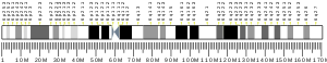
The major histocompatibility complex (MHC) is a large locus on vertebrate DNA containing a set of closely linked polymorphic genes that code for cell surface proteins essential for the adaptive immune system. These cell surface proteins are called MHC molecules.
An antigen-presenting cell (APC) or accessory cell is a cell that displays antigen bound by major histocompatibility complex (MHC) proteins on its surface; this process is known as antigen presentation. T cells may recognize these complexes using their T cell receptors (TCRs). APCs process antigens and present them to T-cells.
MHC class I molecules are one of two primary classes of major histocompatibility complex (MHC) molecules and are found on the cell surface of all nucleated cells in the bodies of vertebrates. They also occur on platelets, but not on red blood cells. Their function is to display peptide fragments of proteins from within the cell to cytotoxic T cells; this will trigger an immediate response from the immune system against a particular non-self antigen displayed with the help of an MHC class I protein. Because MHC class I molecules present peptides derived from cytosolic proteins, the pathway of MHC class I presentation is often called cytosolic or endogenous pathway.
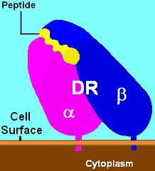
HLA-DR is an MHC class II cell surface receptor encoded by the human leukocyte antigen complex on chromosome 6 region 6p21.31. The complex of HLA-DR and peptide, generally between 9 and 30 amino acids in length, constitutes a ligand for the T-cell receptor (TCR). HLA were originally defined as cell surface antigens that mediate graft-versus-host disease. Identification of these antigens has led to greater success and longevity in organ transplant.
HLA class II histocompatibility antigen, DR alpha chain is a protein that in humans is encoded by the HLA-DRA gene. HLA-DRA encodes the alpha subunit of HLA-DR. Unlike the alpha chains of other Human MHC class II molecules, the alpha subunit is practically invariable. However it can pair with, in any individual, the beta chain from 3 different DR beta loci, DRB1, and two of any DRB3, DRB4, or DRB5 alleles. Thus there is the potential that any given individual can form 4 different HLA-DR isoforms.
Antigen presentation is a vital immune process that is essential for T cell immune response triggering. Because T cells recognize only fragmented antigens displayed on cell surfaces, antigen processing must occur before the antigen fragment can be recognized by a T-cell receptor. Specifically, the fragment, bound to the major histocompatibility complex (MHC), is transported to the surface of the cell, a process known as presentation. If there has been an infection with viruses or bacteria, the cell will present an endogenous or exogenous peptide fragment derived from the antigen by MHC molecules. There are two types of MHC molecules which differ in the behaviour of the antigens: MHC class I molecules (MHC-I) bind peptides from the cell cytosol, while peptides generated in the endocytic vesicles after internalisation are bound to MHC class II (MHC-II). Cellular membranes separate these two cellular environments - intracellular and extracellular. Each T cell can only recognize tens to hundreds of copies of a unique sequence of a single peptide among thousands of other peptides presented on the same cell, because an MHC molecule in one cell can bind to quite a large range of peptides. Predicting which antigens will be presented to the immune system by a certain MHC/HLA type is difficult, but the technology involved is improving.
MHC Class II molecules are a class of major histocompatibility complex (MHC) molecules normally found only on professional antigen-presenting cells such as dendritic cells, mononuclear phagocytes, some endothelial cells, thymic epithelial cells, and B cells. These cells are important in initiating immune responses.
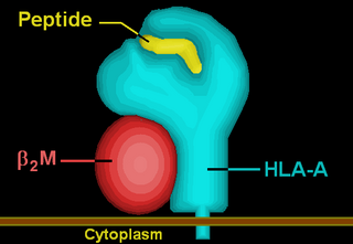
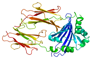
HLA-A is a group of human leukocyte antigens (HLA) that are encoded by the HLA-A locus, which is located at human chromosome 6p21.3. HLA is a major histocompatibility complex (MHC) antigen specific to humans. HLA-A is one of three major types of human MHC class I transmembrane proteins. The others are HLA-B and HLA-C. The protein is a heterodimer, and is composed of a heavy α chain and smaller β chain. The α chain is encoded by a variant HLA-A gene, and the β chain (β2-microglobulin) is an invariant β2 microglobulin molecule. The β2 microglobulin protein is encoded by the B2M gene, which is located at chromosome 15q21.1 in humans.
HLA class I histocompatibility antigen, alpha chain E (HLA-E) also known as MHC class I antigen E is a protein that in humans is encoded by the HLA-E gene. The human HLA-E is a non-classical MHC class I molecule that is characterized by a limited polymorphism and a lower cell surface expression than its classical paralogues. The functional homolog in mice is called Qa-1b, officially known as H2-T23.
CD94, also known as killer cell lectin-like receptor subfamily D, member 1 (KLRD1) is a human gene.
Minor histocompatibility antigen are peptides presented on the cellular surface of donated organs that are known to give an immunological response in some organ transplants. They cause problems of rejection less frequently than those of the major histocompatibility complex (MHC). Minor histocompatibility antigens (MiHAs) are diverse, short segments of proteins and are referred to as peptides. These peptides are normally around 9-12 amino acids in length and are bound to both the major histocompatibility complex (MHC) class I and class II proteins. Peptide sequences can differ among individuals and these differences arise from SNPs in the coding region of genes, gene deletions, frameshift mutations, or insertions. About a third of the characterized MiHAs come from the Y chromosome. Prior to becoming a short peptide sequence, the proteins expressed by these polymorphic or diverse genes need to be digested in the proteasome into shorter peptides. These endogenous or self peptides are then transported into the endoplasmic reticulum with a peptide transporter pump called TAP where they encounter and bind to the MHC class I molecule. This contrasts with MHC class II molecules's antigens which are peptides derived from phagocytosis/endocytosis and molecular degradation of non-self entities' proteins, usually by antigen-presenting cells. MiHA antigens are either ubiquitously expressed in most tissue like skin and intestines or restrictively expressed in the immune cells.
HLA-DM is an intracellular protein involved in the mechanism of antigen presentation on antigen presenting cells (APCs) of the immune system. It does this by assisting in peptide loading of major histocompatibility complex (MHC) class II membrane-bound proteins. HLA-DM is encoded by the genes HLA-DMA and HLA-DMB.
Major histocompatibility complex, class II, DR beta 4, also known as HLA-DRB4, is a human gene.
HLA-G histocompatibility antigen, class I, G, also known as human leukocyte antigen G (HLA-G), is a protein that in humans is encoded by the HLA-G gene.
HLA class II histocompatibility antigen gamma chain also known as HLA-DR antigens-associated invariant chain or CD74, is a protein that in humans is encoded by the CD74 gene. The invariant chain is a polypeptide which plays a critical role in antigen presentation. It is involved in the formation and transport of MHC class II peptide complexes for the generation of CD4+ T cell responses. The cell surface form of the invariant chain is known as CD74. CD74 is a cell surface receptor for the cytokine macrophage migration inhibitory factor (MIF).
MHC class I polypeptide–related sequence A (MICA) is a highly polymorphic cell surface glycoprotein encoded by the MICA gene located within MHC locus. MICA is related to MHC class I and it has similar domain structure, however, it is not associated with β2-microglobulin nor binds peptides as conventional MHC class I molecules do. MICA rather functions as a stress-induced ligand (as a danger signal) for integral membrane protein receptor NKG2D ("natural-killer group 2, member D"). MICA is broadly recognized by NK cells, γδ T cells, and CD8+ αβ T cells which carry NKG2D receptor on their cell surface and which are activated via this interaction.
HLA class II histocompatibility antigen, DRB3-1 beta chain is a protein that in humans is encoded by the HLA-DRB3 gene.
HLA class II histocompatibility antigen, DM beta chain is a protein that in humans is encoded by the HLA-DMB gene.
HLA class II histocompatibility antigen, DM alpha chain is a protein that in humans is encoded by the HLA-DMA gene.
HLA class II histocompatibility antigen, DO beta chain is a protein that in humans is encoded by the HLA-DOB gene.