CD36

Last updated
CD36
Identifiers
Aliases CD36 , BDPLT10, CHDS7, FAT, GP3B, GP4, GPIV, PASIV, SCARB3, CD36 molecule
External IDs OMIM: 173510 MGI: 107899 HomoloGene: 73871 GeneCards: CD36
Orthologs
SpeciesHumanMouse
Entrez
Ensembl
UniProt
RefSeq (mRNA)

NM_001159555
NM_001159556
NM_001159557
NM_001159558
NM_007643

RefSeq (protein)

NP_001153027
NP_001153028
NP_001153029
NP_001153030
NP_031669

Location (UCSC) Chr 7: 80.37 – 80.68 Mb Chr 5: 17.99 – 18.09 Mb
PubMed search [3] [4]
Wikidata
View/Edit Human View/Edit Mouse

CD36 (cluster of differentiation 36), also known as platelet glycoprotein 4, fatty acid translocase (FAT), scavenger receptor class B member 3 (SCARB3), and glycoproteins 88 (GP88), IIIb (GPIIIB), or IV (GPIV) is a protein that in humans is encoded by the CD36 gene. The CD36 antigen is an integral membrane protein found on the surface of many cell types in vertebrate animals. It imports fatty acids inside cells and is a member of the class B scavenger receptor family of cell surface proteins. CD36 binds many ligands including collagen, [5] thrombospondin, [6] erythrocytes parasitized with Plasmodium falciparum , [7] oxidized low density lipoprotein, [8] [9] native lipoproteins, [10] oxidized phospholipids, [11] and long-chain fatty acids. [12]

Work in genetically modified rodents suggest a role for CD36 in fatty acid metabolism, [13] [14] [15] heart disease, [16] taste, [17] [18] [19] and dietary fat processing in the intestine. [20] It may be involved in glucose intolerance, atherosclerosis, arterial hypertension, diabetes, cardiomyopathy, Alzheimer's disease and various cancers, mostly of epithelial origin (breast, prostate, ovary, and colon) and also for hepatic carcinoma and gliomas. [21] [22] [23]

Structure

Primary

In humans, rats and mice, CD36 consists of 472 amino acids with a predicted molecular weight of approximately 53,000 Da. However, CD36 is extensively glycosylated and has an apparent molecular weight of 88,000 Da as determined by SDS polyacrylamide gel electrophoresis. [24]

Tertiary

Using Kyte-Doolittle analysis, [25] the amino acid sequence of CD36 predicts a hydrophobic region near each end of the protein large enough to span cellular membranes. Based on this notion and the observation that CD36 is found on the surface of cells, CD36 is thought to have a 'hairpin-like' structure with α-helices at the C- and N- termini projecting through the membrane and a larger extracellular loop (Fig. 1). This topology is supported by transfection experiments in cultured cells using deletion mutants of CD36. [26] [27]

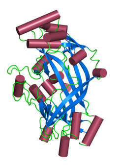
Based on the crystal structure of the homologous SCARB2, a model of the extracellular domain of CD36 has been produced. [28] Like SCARB2, CD36 is proposed to contain an antiparallel β-barrel core with many short α-helices adorning it. The structure is predicted to contain a hydrophobic transport tunnel. Disulfide linkages between 4 of the 6 cysteine residues in the extracellular loop are required for efficient intracellular processing and transport of CD36 to the plasma membrane. [29] It is not clear what role these linkages play on the function of the mature CD36 protein on the cell surface.

Posttranslational modification

Besides glycosylation, additional post-translational modifications have been reported for CD36. CD36 is modified with 4 palmitoyl chains, 2 on each of the two intracellular domains. [27] The function of these lipid modifications is currently unknown but they likely promote the association of CD36 with the membrane and possibly lipid rafts which appear to be important for some CD36 functions. [30] [31] CD36 could be also phosphorylated at Y62, T92, T323, [32] ubiquitinated at K56, K469, K472 and acetylated at K52, K56, K166, K231, K394, K398, K403. [33] [34] [35]

Protein-protein interactions

In the absence of ligand, membrane bound CD36 exists primarily in a monomeric state. However exposure to the thrombospondin ligand causes CD36 to dimerize. This dimerization has been proposed to play an important role in CD36 signal transduction. [36]

Genetics

In humans, the gene is located on the long arm of chromosome 7 at band 11.2 (7q11.2 [37] ) and is encoded by 15 exons that extend over more than 32 kilobases. Both the 5' and the 3' untranslated regions contain introns: the 5' with two and the 3' one. Exons 1, 2 and first 89 nucleotides of exon 3 and as well as exon 15 are non-coding. Exon 3 contains encodes the N-terminal cytoplasmic and transmembrane domains. The C-terminal cytoplasmic and transmembrane regions is encoded by exon 14. The extracellular domain is encoded by the central 11 exons. Alternative splicing of the untranslated regions gives rise to at least two mRNA species.

The transcription initiation site of the CD36 gene has been mapped to 289 nucleotides upstream from the translational start codon and a TATA box and several putative cis regulatory regions lie further 5'. A binding site for PEBP2/CBF factors has been identified between -158 and -90 and disruption of this site reduces expression. The gene is the transcriptional control of the nuclear receptor PPAR/RXR heterodimer (Peroxisome proliferator-activated receptorRetinoid X receptor) and gene expression can be up regulated using synthetic and natural ligands for PPAR and RXR, including the thiazolidinedione class of anti-diabetic drugs and the vitamin A metabolite 9-cis-retinoic acid respectively.

Tissue distribution

CD36 is found on platelets, erythrocytes, monocytes, differentiated adipocytes, skeletal muscle, mammary epithelial cells, spleen cells and some skin microdermal endothelial cells.

Function

The protein itself belongs to the class B scavenger receptor family which includes receptors for selective cholesteryl ester uptake, scavenger receptor class B type I (SR-BI) and lysosomal integral membrane protein II (LIMP-II).

CD36 interacts with a number of ligands, including collagen types I and IV, thrombospondin, erythrocytes parasitized with Plasmodium falciparum , platelet-agglutinating protein p37, oxidized low density lipoprotein and long-chain fatty acids. [38]

On macrophages CD36 forms part of a non-opsonic receptor (the scavenger receptor CD36/alpha-v beta-3 complex) and is involved in phagocytosis. [39]

CD36 has also been implicated in hemostasis, thrombosis, malaria, inflammation, lipid metabolism and atherogenesis. [40]

On binding a ligand the protein and ligand are internalized. This internalization is independent of macropinocytosis and occurs by an actin dependent mechanism requiring the activation Src-family kinases, JNK and Rho-family GTPases. [41] Unlike macropinocytosis this process is not affected by inhibitors of phosphatidylinositol 3-kinase or Na+/H+ exchange.

CD36 ligands have also been shown to promote sterile inflammation through assembly of a Toll-like receptor 4 and 6 heterodimer. [42]

Recently, CD36 was linked to store-operated calcium flux, phospholipase A2 activation, and production of prostaglandin E2 [43]

CD36 function in long-chain fatty acid uptake and signaling can be irreversibly inhibited by sulfo-N-succinimidyl oleate (SSO), which binds lysine 164 within a hydrophobic pocket shared by several CD36 ligands, e.g. fatty acid and oxLDL. [34] Recent research concluded that CD36 is involved in the fat taste transduction (oleogustus).

Clinical significance

Malaria

Infections with the human malaria parasite Plasmodium falciparum are characterized by sequestration of erythrocytes infected with mature forms of the parasite and CD36 has been shown to be a major sequestration receptor on microvascular endothelial cells. Parasitised erythrocytes adhere to endothelium at the trophozoite/schizonts stage simultaneous with the appearance of the var gene product (erythrocyte membrane protein 1) on the erythrocyte surface. The appearance of Plasmodium falciparum erythrocyte membrane protein 1 (PfEMP1) on the erythrocyte surface is a temperature dependent phenomenon which is due to increased protein trafficking to the erythrocyte surface at the raised temperature. PfEMP1 can bind other endothelial receptors - thrombospondin (TSP) and intercellular adhesion molecule 1 (ICAM-1) – in addition to CD36 - and genes other than PfEMP1 also bind to CD36: cytoadherence linked protein (clag) and sequestrin. The PfEMP1 binding site on CD36 is known to be located on exon 5.

CD36 on the surface of the platelets has been shown to be involved in adherence but direct adherence to the endothelium by the infected erythrocytes also occurs. Autoaggregation of infected erythrocytes by platelets has been shown to correlate with severe malaria and cerebral malaria in particular and antiplatelet antibodies may offer some protection.

Several lines of evidence suggest that mutations in CD36 are protective against malaria: mutations in the promoters and within introns and in exon 5 reduce the risk of severe malaria. Gene diversity studies suggest there has been positive selection on this gene presumably due to malarial selection pressure. Dissenting reports are also known suggesting that CD36 is not the sole determinant of severe malaria. In addition a role for CD36 has been found in the clearance of gametocytes (stages I and II).

CD36 has been shown to have a role in the innate immune response to malaria in mouse models. [44] Compared with wild type mice CD36 (-/-) mice the cytokine induction response and parasite clearance were impaired. Earlier peak parasitemias, higher parasite densities and higher mortality were noted. It is thought that CD36 is involved in the Plasmodium falciparum glycophosphatidylinositol (PfGPI) induced MAPK activation and proinflammatory cytokine secretion. When macrophages were exposed to PfGPI the proteins ERK1/2, JNK, p38, and c-Jun became phosphorylated. All these proteins are involved as secondary messengers in the immune response. These responses were blunted in the CD36 (-/-) mice. Also in the CD36 (-/-) macrophages secreted significantly less TNF-alpha on exposure to PfGPI. Work is ongoing to determine how these exactly how these responses provide protection against malaria.

CD36 deficiency and alloimmune thrombocytopenia

CD36 is also known as glycoprotein IV (gpIV) or glycoprotein IIIb (gpIIIb) in platelets and gives rise to the Naka antigen. The Naka null phenotype is found in 0.3% of Caucasians and appears to be asymptomatic. The null phenotype is more common in African (2.5%), Japanese, and other Asian populations (5-11%).

Mutations in the human CD36 gene were first identified in a patient who, despite multiple platelet transfusions, continued to exhibit low platelet levels. [45] [46] This condition is known as refractoriness to platelet transfusion. Subsequent studies have shown that CD36 found on the surface of platelets. This antigen is recognized by the monoclonal antibodies (MAbs) OKM5 and OKM8. It is bound by the Plasmodium falciparum protein sequestrin. [47]

Depending on the nature of the mutation in codon 90 CD36 may be absent either on both platelets and monocytes (type 1) or platelets alone (type 2). Type 2 has been divided into two subtypes - a and b. Deficiency restricted to the platelets alone is known as type 2a; if CD36 is also absent from the erythroblasts the phenotype is classified as type 2b. [48] The molecular basis is known for some cases: T1264G in both Kenyans and Gambians; C478T (50%), 539 deletion of AC and 1159 insertion of an A, 1438-1449 deletion and a combined 839-841 deletion GAG and insertion of AAAAC in Japanese.

In a study of 827 apparently healthy Japanese volunteers, type I and II deficiencies were found in 8 (1.0%) and 48 (5.8%) respectively. [49] In 1127 healthy French blood donors (almost all of whom were white Europeans) no CD36 deficiency was found. [50] In a second group only 1 of 301 white test subjects was found to be CD36 deficient. 16 of the 206 sub-Saharan black Africans and 1 of 148 black Caribbeans were found to be CD36 -ve. Three of 13 CD36 -ve persons examined had anti CD36 antibodies. In a group of 250 black American blood donors 6 (2.4%) were found to be Naka antigen negative. [51]

CD36 deficiency may be a cause of post transfusion purpura. [52]

Blood pressure

Below normal levels of CD36 expression in the kidneys has been implicated as a genetic risk factor for hypertension (high blood pressure). [53]

Fatty acid uptake

An association with myocardial fatty acid uptake in humans has been noted. [54] The data suggest a link between hypertrophic cardiomyopathy and CD36 but this needs to be confirmed.

Tuberculosis

RNAi screening in a Drosophila model has revealed that a member of the CD36 family is required for phagocytosis of Mycobacterium tuberculosis into macrophage phagosomes. [55]

Toxoplasmosis

Avirulent strains of Toxoplasma gondii bind to CD36 but virulent parasites fail to engage CD36. In mice, CD36 is required for disease tolerance but not for the development of immunity or resistance. [56]

Obesity

CD36's association with the ability to taste fats has made it a target for various studies regarding obesity and alteration of lipid tasting. CD36 mRNA expression was found to be reduced in taste bud cells (TBC) of obese sand rats (P. obesus) compared to lean controls, implicating an association between CD36 and obesity. [57] Although actual levels of CD36 protein were not different between the obese and control rat cells, Abdoul-Azize et al. hypothesize that the physical distribution of CD36 could differ in obese rat cells. [57] Changes in calcium mediation have been associated with CD36 and obesity as well. Taste bud cells (more specifically, cells from the circumvallate papillae) containing CD36 that were isolated from obese mice exhibited a significantly smaller increase in calcium after fatty acid stimulation when compared to control mice: [58] CD36 associated calcium regulation is impaired when mice are made to be obese (but not in normal weight mice), and this could be a mechanism contributing to behavior changes in the obese mice, such as decreased lipid taste sensitivity and decreased attraction to fats. [58]

There has been some investigation into human CD36 as well. A study examined oral detection of fat in obese subjects with genetic bases for high, medium, and low expression of the CD36 receptor. Those subjects with high CD36 expression were eight times more sensitive to certain fats (oleic acid and triolein) than the subjects with low CD36 expression. [18] Those subjects with an intermediate amount of CD36 expression were sensitive to fat at a level between the high and low groups. [18] This study demonstrates that there is a significant relationship between oral fat sensitivity and the amount of CD36 receptor expression, but further investigation into CD36 could be useful for learning more about lipid tasting in the context of obesity, as CD36 may be a target for therapies in the future.

Establishment of cellular senescence

Upregulation of CD36 could contribute to membrane remodeling during senescence. [59] In response to various senescence‐inducing stimuli, CD36 stimulate NF-κB‐dependent inflammatory cytokine and chemokine production, a phenomenon known as the senescence‐associated secretory phenotype (SASP). [60] This secretory molecule production leads to the onset of a comprehensive senescent cell fate. Reducing the burden of senescent cells, or reducing their inflammatory secretome through CD36 neutralization, accelerates regeneration in young and old mice. [61]

Cancer

CD36 plays a role in the regulation of angiogenesis, which may be a therapeutic strategy for controlling the spread of cancer. [62] Some data from in vitro and animal studies suggested that fatty acid uptake through CD36 may promote cancer cell migration and proliferation in hepatocellular carcinoma, glioblastoma, and potentially other cancers; there was limited data from observational studies in people that low CD36 may correlate with a slightly better outcome in glioblastoma. [63]

Interactions

CD36 has been shown to interact with FYN. [64] [65]

CD36 family
4f7b 1.png
Structure of Limp-II. PDB entry 4f7b
Identifiers
SymbolCD36
Pfam PF01130
InterPro IPR002159
Available protein structures:
Pfam   structures / ECOD  
PDB RCSB PDB; PDBe; PDBj
PDBsum structure summary

Other human scavenger receptors related to CD36 are SCARB1 and SCARB2 proteins.

See also

Related Research Articles

<span class="mw-page-title-main">Lipolysis</span> Metabolism involving breakdown of lipids

Lipolysis is the metabolic pathway through which lipid triglycerides are hydrolyzed into a glycerol and free fatty acids. It is used to mobilize stored energy during fasting or exercise, and usually occurs in fat adipocytes. The most important regulatory hormone in lipolysis is insulin; lipolysis can only occur when insulin action falls to low levels, as occurs during fasting. Other hormones that affect lipolysis include glucagon, epinephrine, norepinephrine, growth hormone, atrial natriuretic peptide, brain natriuretic peptide, and cortisol.

<i>Plasmodium falciparum</i> Protozoan species of malaria parasite

Plasmodium falciparum is a unicellular protozoan parasite of humans, and the deadliest species of Plasmodium that causes malaria in humans. The parasite is transmitted through the bite of a female Anopheles mosquito and causes the disease's most dangerous form, falciparum malaria. It is responsible for around 50% of all malaria cases. P. falciparum is therefore regarded as the deadliest parasite in humans. It is also associated with the development of blood cancer and is classified as a Group 2A (probable) carcinogen.

Glycophorin C plays a functionally important role in maintaining erythrocyte shape and regulating membrane material properties, possibly through its interaction with protein 4.1. Moreover, it has previously been shown that membranes deficient in protein 4.1 exhibit decreased content of glycophorin C. It is also an integral membrane protein of the erythrocyte and acts as the receptor for the Plasmodium falciparum protein PfEBP-2.

<span class="mw-page-title-main">Duffy antigen system</span> Human blood group classification

Duffy antigen/chemokine receptor (DARC), also known as Fy glycoprotein (FY) or CD234, is a protein that in humans is encoded by the ACKR1 gene.

<span class="mw-page-title-main">Complement receptor 1</span> Mammalian protein found in Homo sapiens

Complement receptor type 1 (CR1) also known as C3b/C4b receptor or CD35 is a protein that in humans is encoded by the CR1 gene.

Scavenger receptors are a large and diverse superfamily of cell surface receptors. Its properties were first recorded in 1970 by Drs. Brown and Goldstein, with the defining property being the ability to bind and remove modified low density lipoproteins (LDL). Today scavenger receptors are known to be involved in a wide range of processes, such as: homeostasis, apoptosis, inflammatory diseases and pathogen clearance. Scavenger receptors are mainly found on myeloid cells and other cells that bind to numerous ligands, primarily endogenous and modified host-molecules together with pathogen-associated molecular patterns(PAMPs), and remove them. The Kupffer cells in the liver are particularly rich in scavenger receptors, includes SR-A I, SR-A II, and MARCO.

<span class="mw-page-title-main">Phospholipid scramblase</span> Protein

Scramblase is a protein responsible for the translocation of phospholipids between the two monolayers of a lipid bilayer of a cell membrane. In humans, phospholipid scramblases (PLSCRs) constitute a family of five homologous proteins that are named as hPLSCR1–hPLSCR5. Scramblases are members of the general family of transmembrane lipid transporters known as flippases. Scramblases are distinct from flippases and floppases. Scramblases, flippases, and floppases are three different types of enzymatic groups of phospholipid transportation enzymes. The inner-leaflet, facing the inside of the cell, contains negatively charged amino-phospholipids and phosphatidylethanolamine. The outer-leaflet, facing the outside environment, contains phosphatidylcholine and sphingomyelin. Scramblase is an enzyme, present in the cell membrane, that can transport (scramble) the negatively charged phospholipids from the inner-leaflet to the outer-leaflet, and vice versa.

<span class="mw-page-title-main">VLDL receptor</span> Protein-coding gene in the species Homo sapiens

The very-low-density-lipoprotein receptor (VLDLR) is a transmembrane lipoprotein receptor of the low-density-lipoprotein (LDL) receptor family. VLDLR shows considerable homology with the members of this lineage. Discovered in 1992 by T. Yamamoto, VLDLR is widely distributed throughout the tissues of the body, including the heart, skeletal muscle, adipose tissue, and the brain, but is absent from the liver. This receptor has an important role in cholesterol uptake, metabolism of apolipoprotein E-containing triacylglycerol-rich lipoproteins, and neuronal migration in the developing brain. In humans, VLDLR is encoded by the VLDLR gene. Mutations of this gene may lead to a variety of symptoms and diseases, which include type I lissencephaly, cerebellar hypoplasia, and atherosclerosis.

<span class="mw-page-title-main">Lipid signaling</span> Biological signaling using lipid molecules

Lipid signaling, broadly defined, refers to any biological cell signaling event involving a lipid messenger that binds a protein target, such as a receptor, kinase or phosphatase, which in turn mediate the effects of these lipids on specific cellular responses. Lipid signaling is thought to be qualitatively different from other classical signaling paradigms because lipids can freely diffuse through membranes. One consequence of this is that lipid messengers cannot be stored in vesicles prior to release and so are often biosynthesized "on demand" at their intended site of action. As such, many lipid signaling molecules cannot circulate freely in solution but, rather, exist bound to special carrier proteins in serum.

An apicoplast is a derived non-photosynthetic plastid found in most Apicomplexa, including Toxoplasma gondii, and Plasmodium falciparum and other Plasmodium spp., but not in others such as Cryptosporidium. It originated from algae through secondary endosymbiosis; there is debate as to whether this was a green or red alga. The apicoplast is surrounded by four membranes within the outermost part of the endomembrane system. The apicoplast hosts important metabolic pathways like fatty acid synthesis, isoprenoid precursor synthesis and parts of the heme biosynthetic pathway.

<span class="mw-page-title-main">Basigin</span> Mammalian protein found in Homo sapiens

Basigin (BSG) also known as extracellular matrix metalloproteinase inducer (EMMPRIN) or cluster of differentiation 147 (CD147) is a protein that in humans is encoded by the BSG gene. This protein is a determinant for the Ok blood group system. There are three known antigens in the Ok system; the most common being Oka, OK2 and OK3. Basigin has been shown to be an essential receptor on red blood cells for the human malaria parasite, Plasmodium falciparum. The common isoform of basigin (basigin-2) has two immunoglobulin domains, and the extended form basigin-1 has three.

<span class="mw-page-title-main">Taste receptor</span> Type of cellular receptor that facilitates taste

A taste receptor or tastant is a type of cellular receptor which facilitates the sensation of taste. When food or other substances enter the mouth, molecules interact with saliva and are bound to taste receptors in the oral cavity and other locations. Molecules which give a sensation of taste are considered "sapid".

Blood lipids are lipids in the blood, either free or bound to other molecules. They are mostly transported in a phospholipid capsule, and the type of protein embedded in this outer shell determines the fate of the particle and its influence on metabolism. Examples of these lipids include cholesterol and triglycerides. The concentration of blood lipids depends on intake and excretion from the intestine, and uptake and secretion from cells. Hyperlipidemia is the presence of elevated or abnormal levels of lipids and/or lipoproteins in the blood, and is a major risk factor for cardiovascular disease.

<span class="mw-page-title-main">Free fatty acid receptor 4</span> Protein-coding gene in the species Homo sapiens

Free Fatty acid receptor 4 (FFAR4), also termed G-protein coupled receptor 120 (GPR120), is a protein that in humans is encoded by the FFAR4 gene. This gene is located on the long arm of chromosome 10 at position 23.33. G protein-coupled receptors reside on their parent cells' surface membranes, bind any one of the specific set of ligands that they recognize, and thereby are activated to trigger certain responses in their parent cells. FFAR4 is a rhodopsin-like GPR in the broad family of GPRs which in humans are encoded by more than 800 different genes. It is also a member of a small family of structurally and functionally related GPRs that include at least three other free fatty acid receptors (FFARs) viz., FFAR1, FFAR2, and FFAR3. These four FFARs bind and thereby are activated by certain fatty acids.

<span class="mw-page-title-main">FABP1</span> Protein-coding gene in the species Homo sapiens

FABP1 is a human gene coding for the protein product FABP1. It is also frequently known as liver-type fatty acid-binding protein (LFABP).

Human genetic resistance to malaria refers to inherited changes in the DNA of humans which increase resistance to malaria and result in increased survival of individuals with those genetic changes. The existence of these genotypes is likely due to evolutionary pressure exerted by parasites of the genus Plasmodium which cause malaria. Since malaria infects red blood cells, these genetic changes are most common alterations to molecules essential for red blood cell function, such as hemoglobin or other cellular proteins or enzymes of red blood cells. These alterations generally protect red blood cells from invasion by Plasmodium parasites or replication of parasites within the red blood cell.

CD36 antigen is a transmembrane, highly glycosylated, glycoprotein expressed by monocytes, macrophages, platelets, microvascular endothelial cells and adipose tissues. CD36 recognises oxidized low density lipoprotein, long chain fatty acids, anionic phospholipids, collagen types I, IV and V, thrombospondin and Plasmodium falciparum infected erythrocytes.

<span class="mw-page-title-main">Duffy binding proteins</span>

In molecular biology, Duffy binding proteins are found in Plasmodium. Plasmodium vivax and Plasmodium knowlesi merozoites invade Homo sapiens erythrocytes that express Duffy blood group surface determinants. The Duffy receptor family is localised in micronemes, an organelle found in all organisms of the phylum Apicomplexa.

Russell J. Howard is an Australian-born executive, entrepreneur and scientist. He was a pioneer in the fields of molecular parasitology, especially malaria, and in leading the commercialisation of one of the most important methods used widely today in molecular biology today called “DNA shuffling" or "Molecular breeding", a form of "Directed evolution".

Plasmodium falciparum erythrocyte membrane protein 1 (PfEMP1) is a family of proteins present on the membrane surface of red blood cells that are infected by the malarial parasite Plasmodium falciparum. PfEMP1 is synthesized during the parasite's blood stage inside the RBC, during which the clinical symptoms of falciparum malaria are manifested. Acting as both an antigen and adhesion protein, it is thought to play a key role in the high level of virulence associated with P. falciparum. It was discovered in 1984 when it was reported that infected RBCs had unusually large-sized cell membrane proteins, and these proteins had antibody-binding (antigenic) properties. An elusive protein, its chemical structure and molecular properties were revealed only after a decade, in 1995. It is now established that there is not one but a large family of PfEMP1 proteins, genetically regulated (encoded) by a group of about 60 genes called var. Each P. falciparum is able to switch on and off specific var genes to produce a functionally different protein, thereby evading the host's immune system. RBCs carrying PfEMP1 on their surface stick to endothelial cells, which facilitates further binding with uninfected RBCs, ultimately helping the parasite to both spread to other RBCs as well as bringing about the fatal symptoms of P. falciparum malaria.

References

  1. 1 2 3 GRCh38: Ensembl release 89: ENSG00000135218 - Ensembl, May 2017
  2. 1 2 3 GRCm38: Ensembl release 89: ENSMUSG00000002944 - Ensembl, May 2017
  3. "Human PubMed Reference:". National Center for Biotechnology Information, U.S. National Library of Medicine.
  4. "Mouse PubMed Reference:". National Center for Biotechnology Information, U.S. National Library of Medicine.
  5. Tandon NN, Kralisz U, Jamieson GA (May 1989). "Identification of glycoprotein IV (CD36) as a primary receptor for platelet-collagen adhesion". The Journal of Biological Chemistry. 264 (13): 7576–83. doi: 10.1016/S0021-9258(18)83273-2 . PMID   2468670.
  6. Silverstein RL, Baird M, Lo SK, Yesner LM (August 1992). "Sense and antisense cDNA transfection of CD36 (glycoprotein IV) in melanoma cells. Role of CD36 as a thrombospondin receptor". The Journal of Biological Chemistry. 267 (23): 16607–12. doi: 10.1016/S0021-9258(18)42046-7 . PMID   1379600.
  7. Oquendo P, Hundt E, Lawler J, Seed B (July 1989). "CD36 directly mediates cytoadherence of Plasmodium falciparum parasitized erythrocytes". Cell. 58 (1): 95–101. doi:10.1016/0092-8674(89)90406-6. PMID   2473841. S2CID   22059108.
  8. Endemann G, Stanton LW, Madden KS, Bryant CM, White RT, Protter AA (June 1993). "CD36 is a receptor for oxidized low density lipoprotein". The Journal of Biological Chemistry. 268 (16): 11811–6. doi: 10.1016/S0021-9258(19)50272-1 . PMID   7685021.
  9. Nicholson AC, Frieda S, Pearce A, Silverstein RL (February 1995). "Oxidized LDL binds to CD36 on human monocyte-derived macrophages and transfected cell lines. Evidence implicating the lipid moiety of the lipoprotein as the binding site". Arteriosclerosis, Thrombosis, and Vascular Biology. 15 (2): 269–75. doi: 10.1161/01.ATV.15.2.269 . PMID   7538425.
  10. Calvo D, Gómez-Coronado D, Suárez Y, Lasunción MA, Vega MA (April 1998). "Human CD36 is a high affinity receptor for the native lipoproteins HDL, LDL, and VLDL". Journal of Lipid Research. 39 (4): 777–88. doi: 10.1016/S0022-2275(20)32566-9 . PMID   9555943.[ permanent dead link ]
  11. Podrez EA, Poliakov E, Shen Z, Zhang R, Deng Y, Sun M, et al. (October 2002). "Identification of a novel family of oxidized phospholipids that serve as ligands for the macrophage scavenger receptor CD36". The Journal of Biological Chemistry. 277 (41): 38503–16. doi: 10.1074/jbc.M203318200 . PMID   12105195.
  12. Baillie AG, Coburn CT, Abumrad NA (September 1996). "Reversible binding of long-chain fatty acids to purified FAT, the adipose CD36 homolog". The Journal of Membrane Biology. 153 (1): 75–81. doi:10.1007/s002329900111. PMID   8694909. S2CID   5911289.
  13. Hajri T, Han XX, Bonen A, Abumrad NA (May 2002). "Defective fatty acid uptake modulates insulin responsiveness and metabolic responses to diet in CD36-null mice". The Journal of Clinical Investigation. 109 (10): 1381–9. doi:10.1172/JCI14596. PMC   150975 . PMID   12021254.
  14. Pravenec M, Landa V, Zídek V, Musilová A, Kazdová L, Qi N, et al. (2003). "Transgenic expression of CD36 in the spontaneously hypertensive rat is associated with amelioration of metabolic disturbances but has no effect on hypertension" (PDF). Physiological Research. 52 (6): 681–8. doi:10.33549/physiolres.930380. PMID   14640889.
  15. Mistry JJ, Bowles KM, Rushworth SA (December 2021). "Free fatty-acid transport via CD36 drives β-oxidation-mediated hematopoietic stem cell response to infection". Nature Communications. 12 (1): 7130. Bibcode:2021NatCo..12.7130M. doi:10.1038/s41467-021-27460-9. PMC   8655073 . PMID   34880245.
  16. Febbraio M, Podrez EA, Smith JD, Hajjar DP, Hazen SL, Hoff HF, et al. (April 2000). "Targeted disruption of the class B scavenger receptor CD36 protects against atherosclerotic lesion development in mice". The Journal of Clinical Investigation. 105 (8): 1049–56. doi:10.1172/JCI9259. PMC   300837 . PMID   10772649.
  17. Laugerette F, Passilly-Degrace P, Patris B, Niot I, Febbraio M, Montmayeur JP, Besnard P (November 2005). "CD36 involvement in orosensory detection of dietary lipids, spontaneous fat preference, and digestive secretions". The Journal of Clinical Investigation. 115 (11): 3177–84. doi:10.1172/JCI25299. PMC   1265871 . PMID   16276419.
  18. 1 2 3 Pepino MY, Love-Gregory L, Klein S, Abumrad NA (March 2012). "The fatty acid translocase gene CD36 and lingual lipase influence oral sensitivity to fat in obese subjects". Journal of Lipid Research. 53 (3): 561–6. doi: 10.1194/jlr.M021873 . PMC   3276480 . PMID   22210925.
  19. DiPatrizio NV (September 2014). "Is fat taste ready for primetime?". Physiology & Behavior. 136: 145–54. doi:10.1016/j.physbeh.2014.03.002. PMC   4162865 . PMID   24631296.
  20. Drover VA, Ajmal M, Nassir F, Davidson NO, Nauli AM, Sahoo D, et al. (May 2005). "CD36 deficiency impairs intestinal lipid secretion and clearance of chylomicrons from the blood". The Journal of Clinical Investigation. 115 (5): 1290–7. doi:10.1172/JCI21514. PMC   1074677 . PMID   15841205.
  21. Rać ME, Safranow K, Poncyljusz W (2007). "Molecular basis of human CD36 gene mutations". Molecular Medicine. 13 (5–6): 288–96. doi:10.2119/2006-00088.Rac. PMC   1936231 . PMID   17673938.
  22. Ana-Maria Enciu, Eugen Radu, Ionela Daniela Popescu, Mihail Eugen Hinescu, Laura Cristina Ceafalan (2018). "Targeting CD36 as Biomarker for Metastasis Prognostic: How Far from Translation into Clinical Practice?". BioMed Research International. 2018: 1–12. doi: 10.1155/2018/7801202 . PMC   6057354 . PMID   30069479.
  23. Jingchun Wang, Yongsheng Li (2019). "CD36 tango in cancer: signaling pathways and functions". Theranostics. 9 (17): 4893–490. doi:10.7150/thno.36037. PMC   6691380 . PMID   31410189.
  24. Greenwalt DE, Watt KW, So OY, Jiwani N (July 1990). "PAS IV, an integral membrane protein of mammary epithelial cells, is related to platelet and endothelial cell CD36 (GP IV)". Biochemistry. 29 (30): 7054–9. doi:10.1021/bi00482a015. PMID   1699598.
  25. Kyte J, Doolittle RF (May 1982). "A simple method for displaying the hydropathic character of a protein". Journal of Molecular Biology. 157 (1): 105–32. CiteSeerX   10.1.1.458.454 . doi:10.1016/0022-2836(82)90515-0. PMID   7108955.
  26. Gruarin P, Thorne RF, Dorahy DJ, Burns GF, Sitia R, Alessio M (August 2000). "CD36 is a ditopic glycoprotein with the N-terminal domain implicated in intracellular transport". Biochemical and Biophysical Research Communications. 275 (2): 446–54. doi:10.1006/bbrc.2000.3333. PMID   10964685.
  27. 1 2 Tao N, Wagner SJ, Lublin DM (September 1996). "CD36 is palmitoylated on both N- and C-terminal cytoplasmic tails". The Journal of Biological Chemistry. 271 (37): 22315–20. doi: 10.1074/jbc.271.37.22315 . PMID   8798390.
  28. Neculai D, Schwake M, Ravichandran M, Zunke F, Collins RF, Peters J, et al. (December 2013). "Structure of LIMP-2 provides functional insights with implications for SR-BI and CD36". Nature. 504 (7478): 172–6. Bibcode:2013Natur.504..172N. doi:10.1038/nature12684. PMID   24162852. S2CID   4395239.
  29. Gruarin P, Sitia R, Alessio M (December 1997). "Formation of one or more intrachain disulphide bonds is required for the intracellular processing and transport of CD36". The Biochemical Journal. 328 (2): 635–42. doi:10.1042/bj3280635. PMC   1218965 . PMID   9371725.
  30. Zeng Y, Tao N, Chung KN, Heuser JE, Lublin DM (November 2003). "Endocytosis of oxidized low density lipoprotein through scavenger receptor CD36 utilizes a lipid raft pathway that does not require caveolin-1". The Journal of Biological Chemistry. 278 (46): 45931–6. doi: 10.1074/jbc.M307722200 . PMID   12947091.
  31. Pohl J, Ring A, Korkmaz U, Ehehalt R, Stremmel W (January 2005). "FAT/CD36-mediated long-chain fatty acid uptake in adipocytes requires plasma membrane rafts". Molecular Biology of the Cell. 16 (1): 24–31. doi:10.1091/mbc.E04-07-0616. PMC   539148 . PMID   15496455.
  32. Hornbeck PV, Kornhauser JM, Tkachev S, Zhang B, Skrzypek E, Murray B, Latham V, Sullivan M. "CD36 (human) protein page". PhosphoSitePlus. Cell Signaling Technology, Inc.
  33. Smith J, Su X, El-Maghrabi R, Stahl PD, Abumrad NA (May 2008). "Opposite regulation of CD36 ubiquitination by fatty acids and insulin: effects on fatty acid uptake". The Journal of Biological Chemistry. 283 (20): 13578–85. doi: 10.1074/jbc.M800008200 . PMC   2376227 . PMID   18353783.
  34. 1 2 Kuda O, Pietka TA, Demianova Z, Kudova E, Cvacka J, Kopecky J, Abumrad NA (May 2013). "Sulfo-N-succinimidyl oleate (SSO) inhibits fatty acid uptake and signaling for intracellular calcium via binding CD36 lysine 164: SSO also inhibits oxidized low density lipoprotein uptake by macrophages". The Journal of Biological Chemistry. 288 (22): 15547–55. doi: 10.1074/jbc.M113.473298 . PMC   3668716 . PMID   23603908.
  35. Lundby A, Lage K, Weinert BT, Bekker-Jensen DB, Secher A, Skovgaard T, et al. (August 2012). "Proteomic analysis of lysine acetylation sites in rat tissues reveals organ specificity and subcellular patterns". Cell Reports. 2 (2): 419–31. doi:10.1016/j.celrep.2012.07.006. PMC   4103158 . PMID   22902405.
  36. Daviet L, Malvoisin E, Wild TF, McGregor JL (August 1997). "Thrombospondin induces dimerization of membrane-bound, but not soluble CD36". Thrombosis and Haemostasis. 78 (2): 897–901. doi:10.1055/s-0038-1657649. PMID   9268192. S2CID   43232897.
  37. Fernández-Ruiz E, Armesilla AL, Sánchez-Madrid F, Vega MA (September 1993). "Gene encoding the collagen type I and thrombospondin receptor CD36 is located on chromosome 7q11.2". Genomics. 17 (3): 759–61. doi:10.1006/geno.1993.1401. hdl: 2436/7706 . PMID   7503937.
  38. Armesilla AL, Vega MA (July 1994). "Structural organization of the gene for human CD36 glycoprotein". The Journal of Biological Chemistry. 269 (29): 18985–91. doi: 10.1016/S0021-9258(17)32263-9 . hdl: 2436/7744 . PMID   7518447.
  39. Erdman LK, Cosio G, Helmers AJ, Gowda DC, Grinstein S, Kain KC (November 2009). "CD36 and TLR interactions in inflammation and phagocytosis: implications for malaria". Journal of Immunology. 183 (10): 6452–9. doi:10.4049/jimmunol.0901374. PMC   2853812 . PMID   19864601.
  40. Daviet L, McGregor JL (July 1997). "Vascular biology of CD36: roles of this new adhesion molecule family in different disease states". Thrombosis and Haemostasis. 78 (1): 65–9. doi:10.1055/s-0038-1657502. PMID   9198129. S2CID   21113427.
  41. Collins RF, Touret N, Kuwata H, Tandon NN, Grinstein S, Trimble WS (October 2009). "Uptake of oxidized low density lipoprotein by CD36 occurs by an actin-dependent pathway distinct from macropinocytosis". The Journal of Biological Chemistry. 284 (44): 30288–97. doi: 10.1074/jbc.M109.045104 . PMC   2781584 . PMID   19740737.
  42. Stewart CR, Stuart LM, Wilkinson K, van Gils JM, Deng J, Halle A, et al. (February 2010). "CD36 ligands promote sterile inflammation through assembly of a Toll-like receptor 4 and 6 heterodimer". Nature Immunology. 11 (2): 155–61. doi:10.1038/ni.1836. PMC   2809046 . PMID   20037584.
  43. Kuda O, Jenkins CM, Skinner JR, Moon SH, Su X, Gross RW, Abumrad NA (May 2011). "CD36 protein is involved in store-operated calcium flux, phospholipase A2 activation, and production of prostaglandin E2". The Journal of Biological Chemistry. 286 (20): 17785–95. doi: 10.1074/jbc.M111.232975 . PMC   3093854 . PMID   21454644.
  44. Patel SN, Lu Z, Ayi K, Serghides L, Gowda DC, Kain KC (March 2007). "Disruption of CD36 impairs cytokine response to Plasmodium falciparum glycosylphosphatidylinositol and confers susceptibility to severe and fatal malaria in vivo". Journal of Immunology. 178 (6): 3954–61. doi: 10.4049/jimmunol.178.6.3954 . PMID   17339496.
  45. Ikeda H, Mitani T, Ohnuma M, Haga H, Ohtzuka S, Kato T, et al. (1989). "A new platelet-specific antigen, Naka, involved in the refractoriness of HLA-matched platelet transfusion". Vox Sanguinis. 57 (3): 213–7. doi: 10.1111/j.1423-0410.1989.tb00826.x . PMID   2617957. S2CID   39521299.
  46. Yamamoto N, Ikeda H, Tandon NN, Herman J, Tomiyama Y, Mitani T, et al. (November 1990). "A platelet membrane glycoprotein (GP) deficiency in healthy blood donors: Naka- platelets lack detectable GPIV (CD36)". Blood. 76 (9): 1698–703. doi: 10.1182/blood.V76.9.1698.1698 . PMID   1699620.
  47. Ockenhouse CF, Klotz FW, Tandon NN, Jamieson GA (April 1991). "Sequestrin, a CD36 recognition protein on Plasmodium falciparum malaria-infected erythrocytes identified by anti-idiotype antibodies". Proceedings of the National Academy of Sciences of the United States of America. 88 (8): 3175–9. Bibcode:1991PNAS...88.3175O. doi: 10.1073/pnas.88.8.3175 . PMC   51408 . PMID   1707534.
  48. Toba K, Hanawa H, Watanabe K, Fuse I, Masuko M, Miyajima S, et al. (October 2001). "Erythroid involvement in CD36 deficiency". Experimental Hematology. 29 (10): 1194–200. doi: 10.1016/S0301-472X(01)00691-9 . PMID   11602321.
  49. Yanai H, Chiba H, Fujiwara H, Morimoto M, Abe K, Yoshida S, et al. (September 2000). "Phenotype-genotype correlation in CD36 deficiency types I and II". Thrombosis and Haemostasis. 84 (3): 436–41. doi:10.1055/s-0037-1614041. PMID   11019968. S2CID   42193140.
  50. Lee K, Godeau B, Fromont P, Plonquet A, Debili N, Bachir D, et al. (August 1999). "CD36 deficiency is frequent and can cause platelet immunization in Africans". Transfusion. 39 (8): 873–9. doi: 10.1046/j.1537-2995.1999.39080873.x . PMID   10504124. S2CID   21921171.
  51. Curtis BR, Aster RH (April 1996). "Incidence of the Nak(a)-negative platelet phenotype in African Americans is similar to that of Asians". Transfusion. 36 (4): 331–4. doi:10.1046/j.1537-2995.1996.36496226147.x. PMID   8623134. S2CID   10991605.
  52. Bierling P, Godeau B, Fromont P, Bettaieb A, Debili N, el-Kassar N, et al. (September 1995). "Posttransfusion purpura-like syndrome associated with CD36 (Naka) isoimmunization". Transfusion. 35 (9): 777–82. doi:10.1046/j.1537-2995.1995.35996029165.x. PMID   7570941. S2CID   22706156.
  53. Pravenec M, Churchill PC, Churchill MC, Viklicky O, Kazdova L, Aitman TJ, et al. (August 2008). "Identification of renal Cd36 as a determinant of blood pressure and risk for hypertension". Nature Genetics. 40 (8): 952–4. doi:10.1038/ng.164. PMID   18587397. S2CID   6857655.
  54. Okamoto F, Tanaka T, Sohmiya K, Kawamura K (July 1998). "CD36 abnormality and impaired myocardial long-chain fatty acid uptake in patients with hypertrophic cardiomyopathy". Japanese Circulation Journal. 62 (7): 499–504. doi: 10.1253/jcj.62.499 . PMID   9707006.
  55. Philips JA, Rubin EJ, Perrimon N (August 2005). "Drosophila RNAi screen reveals CD36 family member required for mycobacterial infection". Science. 309 (5738): 1251–3. Bibcode:2005Sci...309.1251P. doi: 10.1126/science.1116006 . PMID   16020694. S2CID   26751583.
  56. Zhao Y, Reyes, J, Rovira-Diaz E, Fox BA, Bzik D, Yap GS (August 2021). "CD36 mediates phagocyte tropism and avirulence of Toxoplasma gondii". Journal of Immunology. 207 (6): 1507–1512. doi:10.4049/jimmunol.2100605. ISSN   0022-1767. PMC   8429199 . PMID   34400524. S2CID   237148810.
  57. 1 2 Abdoul-Azize S, Atek-Mebarki F, Bitam A, Sadou H, Koceïr EA, Khan NA (2013). "Oro-gustatory perception of dietary lipids and calcium signaling in taste bud cells are altered in nutritionally obesity-prone Psammomys obesus". PLOS ONE. 8 (8): e68532. Bibcode:2013PLoSO...868532A. doi: 10.1371/journal.pone.0068532 . PMC   3731325 . PMID   23936306.
  58. 1 2 Chevrot M, Bernard A, Ancel D, Buttet M, Martin C, Abdoul-Azize S, et al. (September 2013). "Obesity alters the gustatory perception of lipids in the mouse: plausible involvement of lingual CD36". Journal of Lipid Research. 54 (9): 2485–94. doi: 10.1194/jlr.M039446 . PMC   3735945 . PMID   23840049.
  59. Saitou M, Lizardo DY, Taskent RO, Millner A, Gokcumen O, Atilla-Gokcumen GE (2018). "An evolutionary transcriptomics approach links CD36 to membrane remodeling in replicative senescence" (PDF). Molecular Omics. Royal Society of Chemistry (RSC). 14 (4): 237–246. bioRxiv   10.1101/294512 . doi:10.1039/c8mo00099a. ISSN   2515-4184. PMID   29974107.
  60. Chong M, et al. (2018). "CD36 initiates the secretory phenotype during the establishment of cellular senescence". EMBO Reports. 19 (6). doi:10.15252/embr.201745274. PMC   5989758 . PMID   29777051.
  61. Moiseeva V, et al. (2022). "Senescence atlas reveals an aged-like inflamed niche that blunts muscle regeneration". Nature. 612 (7941): 169–178. doi:10.1038/s41586-022-05535-x. PMC   9812788 . PMID   36544018.
  62. Ge Y, Elghetany MT (2005). "CD36: a multiligand molecule". Laboratory Hematology. 11 (1): 31–7. doi:10.1532/LH96.04056. PMID   15790550.
  63. Selwan EM, Finicle BT, Kim SM, Edinger AL (April 2016). "Attacking the supply wagons to starve cancer cells to death". FEBS Letters. 590 (7): 885–907. doi:10.1002/1873-3468.12121. PMC   4833639 . PMID   26938658.
  64. Huang MM, Bolen JB, Barnwell JW, Shattil SJ, Brugge JS (September 1991). "Membrane glycoprotein IV (CD36) is physically associated with the Fyn, Lyn, and Yes protein-tyrosine kinases in human platelets". Proceedings of the National Academy of Sciences of the United States of America. 88 (17): 7844–8. Bibcode:1991PNAS...88.7844H. doi: 10.1073/pnas.88.17.7844 . PMC   52400 . PMID   1715582.
  65. Bull HA, Brickell PM, Dowd PM (August 1994). "Src-related protein tyrosine kinases are physically associated with the surface antigen CD36 in human dermal microvascular endothelial cells". FEBS Letters. 351 (1): 41–4. doi:10.1016/0014-5793(94)00814-0. PMID   7521304. S2CID   45071719.

Further reading