Schwannoma

Last updated
Schwannoma
Other namesneurilemoma, [1] :621 neuroma, [2] neurolemoma, [2] Schwann cell tumor [2]
Schwannoma with Antoni A and Antoni B areas.jpg
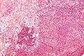
Micrograph of a schwannoma showing both a cellular Antoni A area (top) and a loose paucicellular Antoni B area (bottom). HE stain.
Specialty Neuro-oncology

A schwannoma (or neurilemmoma) is a usually benign nerve sheath tumor composed of Schwann cells, which normally produce the insulating myelin sheath covering peripheral nerves.

Contents

Schwannomas are homogeneous tumors, consisting only of Schwann cells. The tumor cells always stay on the outside of the nerve, but the tumor itself may either push the nerve aside and/or up against a bony structure (thereby possibly causing damage). Schwannomas are relatively slow-growing. For reasons not yet understood, schwannomas are mostly benign and less than 1% become malignant, degenerating into a form of cancer known as neurofibrosarcoma. These masses are generally contained within a capsule, so surgical removal is often successful. [3]

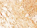
Schwannomas can be associated with neurofibromatosis type II, which may be due to a loss-of-function mutation in the protein merlin. [4] They are universally S-100 positive, which is a marker for cells of neural crest cell origin.

Schwannomas of the head and neck are a fairly common occurrence and can be found incidentally in 3–4% of patients at autopsy. [4] Most common of these is a vestibular schwannoma, a tumor of the vestibulocochlear nerve that may lead to tinnitus and hearing loss on the affected side. Outside the cranial nerves, schwannomas may present on the flexor surfaces of the limbs. Rare occurrences of these tumors in the penis have been documented in the literature. [5]

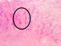
Verocay bodies are seen histologically in schwannomas.

Variants


See also

Related Research Articles

<span class="mw-page-title-main">Neurofibromatosis</span> Three genetic disorders involving benign tumors of the nervous system

Neurofibromatosis (NF) refers to a group of three distinct genetic conditions in which tumors grow in the nervous system. The tumors are non-cancerous (benign) and often involve the skin or surrounding bone. Although symptoms are often mild, each condition presents differently. Neurofibromatosis type I (NF1) is typically characterized by café au lait spots, neurofibromas, scoliosis, and headaches. Neurofibromatosis type II (NF2), on the other hand, may present with early-onset hearing loss, cataracts, tinnitus, difficulty walking or maintain balance, and muscle atrophy. The third type is called schwannomatosis and often presents in early adulthood with widespread pain, numbness, or tingling due to nerve compression.

<span class="mw-page-title-main">Nervous tissue</span> Main component of the nervous system

Nervous tissue, also called neural tissue, is the main tissue component of the nervous system. The nervous system regulates and controls body functions and activity. It consists of two parts: the central nervous system (CNS) comprising the brain and spinal cord, and the peripheral nervous system (PNS) comprising the branching peripheral nerves. It is composed of neurons, also known as nerve cells, which receive and transmit impulses, and neuroglia, also known as glial cells or glia, which assist the propagation of the nerve impulse as well as provide nutrients to the neurons.

<span class="mw-page-title-main">Benign tumor</span> Mass of cells which cannot spread throughout the body

A benign tumor is a mass of cells (tumor) that does not invade neighboring tissue or metastasize. Compared to malignant (cancerous) tumors, benign tumors generally have a slower growth rate. Benign tumors have relatively well differentiated cells. They are often surrounded by an outer surface or stay contained within the epithelium. Common examples of benign tumors include moles and uterine fibroids.

<span class="mw-page-title-main">Neurofibromatosis type I</span> Type of neurofibromatosis disease

Neurofibromatosis type I (NF-1), or von Recklinghausen syndrome, is a complex multi-system human disorder caused by the mutation of neurofibromin 1 (NF-1), a gene on chromosome 17 that is responsible for production of a protein (neurofibromin) which is needed for normal function in many human cell types. NF-1 causes tumors along the nervous system which can grow anywhere on the body. NF-1 is one of the most common genetic disorders and is not limited to any person's race or sex. NF-1 is an autosomal dominant disorder, which means that mutation or deletion of one copy of the NF-1 gene is sufficient for the development of NF-1, although presentation varies widely and is often different even between relatives affected by NF-1.

<span class="mw-page-title-main">Malignant peripheral nerve sheath tumor</span> Cancer of the connective tissue surrounding peripheral nerves

A malignant peripheral nerve sheath tumor (MPNST) is a form of cancer of the connective tissue surrounding peripheral nerves. Given its origin and behavior it is classified as a sarcoma. About half the cases are diagnosed in people with neurofibromatosis; the lifetime risk for an MPNST in patients with neurofibromatosis type 1 is 8–13%. MPNST with rhabdomyoblastomatous component are called malignant triton tumors.

<span class="mw-page-title-main">Neurofibromatosis type II</span> Type of neurofibromatosis disease

Neurofibromatosis type II is a genetic condition that may be inherited or may arise spontaneously, and causes benign tumors of the brain, spinal cord, and peripheral nerves. The types of tumors frequently associated with NF2 include vestibular schwannomas, meningiomas, and ependymomas. The main manifestation of the condition is the development of bilateral benign brain tumors in the nerve sheath of the cranial nerve VIII, which is the "auditory-vestibular nerve" that transmits sensory information from the inner ear to the brain. Besides, other benign brain and spinal tumors occur. Symptoms depend on the presence, localisation and growth of the tumor(s). Many people with this condition also experience vision problems. Neurofibromatosis type II is caused by mutations of the "Merlin" gene, which seems to influence the form and movement of cells. The principal treatments consist of neurosurgical removal of the tumors and surgical treatment of the eye lesions. Historically the underlying disorder has not had any therapy due to the cell function caused by the genetic mutation.

<span class="mw-page-title-main">Neurofibroma</span> Benign nerve-sheath tumor in the peripheral nervous system

A neurofibroma is a benign nerve-sheath tumor in the peripheral nervous system. In 90% of cases, they are found as stand-alone tumors, while the remainder are found in persons with neurofibromatosis type I (NF1), an autosomal-dominant genetically inherited disease. They can result in a range of symptoms from physical disfiguration and pain to cognitive disability.

<span class="mw-page-title-main">Schwannomatosis</span> Rare genetic disorder

Schwannomatosis is an extremely rare genetic disorder closely related to the more-common disorder neurofibromatosis (NF). Originally described in Japanese patients, it consists of multiple cutaneous schwannomas, central nervous system tumors, and other neurological complications, excluding hallmark signs of NF. The exact frequency of schwannomatosis cases is unknown, although some populations have noted frequencies as few as 1 case per 1.7 million people.

<span class="mw-page-title-main">Neurofibromin</span> Mammalian protein found in humans

Neurofibromin (NF-1) is a protein that is encoded in the human by the NF1 gene. NF1 is located on chromosome 17. Neurofibromin, a GTPase-activating protein that negatively regulates RAS/MAPK pathway activity by accelerating the hydrolysis of Ras-bound GTP. NF1 has a high mutation rate and mutations can alter cellular growth control, and neural development, resulting in neurofibromatosis type 1. Symptoms of NF1 include disfiguring cutaneous neurofibromas (CNF), café au lait pigment spots, plexiform neurofibromas (PN), skeletal defects, optic nerve gliomas, life-threatening malignant peripheral nerve sheath tumors (MPNST), pheochromocytoma, attention deficits, learning deficits and other cognitive disabilities.

<span class="mw-page-title-main">Granular cell tumor</span> Medical condition

Granular cell tumor is a tumor that can develop on any skin or mucosal surface, but occurs on the tongue 40% of the time.

A nerve sheath tumor is a type of tumor of the nervous system which is made up primarily of the myelin surrounding nerves. From benign tumors like schwannoma to high grade malignant neoplasms known as malignant peripheral nerve sheath tumors, peripheral nerve sheath tumors include a range of clearly characterized clinicopathologic entities. A peripheral nerve sheath tumor (PNST) is a nerve sheath tumor in the peripheral nervous system. Benign peripheral nerve sheath tumors include schwannomas and neurofibromas.

Malignant triton tumor (MTT) is a relatively rare, aggressive tumor made up of both malignant schwannoma cells and malignant rhabdomyoblasts. It is classified as a malignant peripheral nerve sheath tumor with rhabdomyosarcomatous differentiation.

Plexiform fibrohistiocytic tumor is a rare tumor that arises primarily on the upper extremities of children and young adults.

<span class="mw-page-title-main">WHO classification of tumours of the central nervous system</span>

The WHOclassification of tumours of the central nervous system is a World Health Organization Blue Book that defines, describes and classifies tumours of the central nervous system (CNS).

A rhabdomyoblast is a cell type which is found in some rhabdomyosarcomas. When found histologically, a rhabdomyoblast aids the diagnosis of embryonal, alveolar, spindle cell/sclerosing, and pleomorphic rhabdomyosarcomas; however, in a tumor, expression of the rhabdomyoblast phenotype is not the only factor in diagnosing a rhabdomyosarcoma. Mesenchymal malignancies can exhibit this phenotype as well. Immunohistochemistry techniques allow for the sensitive detection of desmin, vimentin, muscle specific actin, and MyoD1. Similarly the rhabdomyoblast phenotype can be detected morphologically.

Within medical ophthalmology, Intraocular schwannoma, also termed uveal schwannoma, is a type of schwannoma found in the eye. These tumors are almost always benign in nature and while malignant forms have been documented in other areas of the body, this has not been reported in the uveal region. Composed of Schwann cells, these masses are generally slow growing and can be found in the peripheral nerve tract, often around the head and neck.

An intraneural perineurioma is a rare benign tumor within the sheath of a single nerve that grows but typically does not recur or metastasize. These lesions are only composed of perineurial cells, cloned from a single cell. They are distinct from schwannoma and neurofibroma.

<span class="mw-page-title-main">Biphenotypic sinonasal sarcoma</span> Type of nasal cancer

Biphenotypic sinonasal sarcoma is a newly recognized, very rare, low-grade malignant tumor of the nasal cavity, which was formerly probably included in fibrosarcoma and synovial sarcoma cases. It was incorporated into the fourth edition of the World Health Organization Classification of Head and Neck Tumours, published in 2017.

Gastroblastoma is a rare cancer that occurs in the stomach. Only six cases have been reported to date (2017).

<span class="mw-page-title-main">Peripheral nerve tumor</span> Medical condition

Peripheral nerve tumors, also called tumors of peripheral nerves or tumors of the peripheral nervous system, are a diverse category with a range of morphological characteristics and biological potential. They are categorized as either benign or malignant peripheral nerve sheath tumors.

References

  1. James WD, Berger T, Elston D (2006). Andrews' diseases of the skin : clinical dermatology (10th ed.). Philadelphia: Saunders Elsevier. ISBN   978-0-7216-2921-6.
  2. 1 2 3 Rapini RP, Bolognia JL, Jorizzo JL (2007). Dermatology: 2-Volume Set. St. Louis: Mosby. ISBN   978-1-4160-2999-1.
  3. Biswas D, Marnane CN, Mal R, Baldwin D (September 2007). "Extracranial head and neck schwannomas--a 10-year review". Auris, Nasus, Larynx. 34 (3): 353–359. doi:10.1016/j.anl.2007.01.006. PMID   17376620.
  4. 1 2 Hanemann CO, Evans DG (December 2006). "News on the genetics, epidemiology, medical care and translational research of Schwannomas". Journal of Neurology. 253 (12): 1533–1541. doi:10.1007/s00415-006-0347-0. PMID   17219030. S2CID   11854280.
  5. Nguyen AH, Smith ML, Maranda EL, Punnen S (June 2016). "Clinical Features and Treatment of Penile Schwannoma: A Systematic Review". Clinical Genitourinary Cancer. 14 (3): 198–202. doi:10.1016/j.clgc.2015.12.018. PMID   26797586.
  6. Fletcher CD, Davies SE. Benign plexiform (multinodular) schwannoma: a rare tumour unassociated with neurofibromatosis. Histopathology. 1986; 10:971–980. [PubMed: 3096870]
  7. Woodruff JM, Marshall ML, Godwin TA, Funkhouser JW, Thompson NJ, Erlandson RA. Plexiform (multinodular) schwannoma. A tumor simulating the plexiform neurofibroma. Am J Surg Pathol. 1983; 7:691–697. [PubMed: 6638259]
  8. Berg JC, Scheithauer BW, Spinner RJ, Allen CM, Koutlas IG. Plexiform schwannoma: a clinicopathologic overview with emphasis on the head and neck region. Hum Pathol. 2008; 39:633–640. [PubMed: 18439936]
  9. Agaram, N. P.; Prakash, S.; Antonescu, C. R.; "Deep-seated plexiform schwannoma: a pathologic study of 16 cases and comparative analysis with the superficial variety", American Journal of Clinical Pathology , 29:1042–1048, 2005 [PubMed: 16006798]
  10. Hebert-Blouin MN, Amrami KK, Scheithauer BW, Spinner RJ. Multinodular/plexiform (multifascicular) schwannomas of major peripheral nerves: an underrecognized part of the spectrum of schwannomas. J Neurosurg. 2010; 112:372–382. [PubMed: 19499977]
  11. Iwasaki T (2016). "Giant ancient schwannoma of the pleura: Commentary". Lung India. 33 (4): 449–50. doi: 10.4103/0970-2113.184928 . PMC   4948239 . PMID   27578944.
  12. Casadei GP, Scheithauer BW, Hirose T, Manfrini M, Van Houton C, Wood MB. Cellular schwannoma. A clinicopathologic, DNA flow cytometric, and proliferation marker study of 70 patients. Cancer. 1995; 75:1109–1119. [PubMed: 7850709]
  13. Fletcher CD, Davies SE, McKee PH. Cellular schwannoma: a distinct pseudosarcomatous entity. Histopathology. 1987; 11:21–35. [PubMed: 3557324]
  14. Wick, M. R.; Swanson, P. E.; Scheithauer, B. W.; Manivel, J. C.; "Malignant peripheral nerve sheath tumor: An immunohistochemical study of 62 cases", American Journal of Clinical Pathology , 87:425–433, 1987 [PubMed:2435144]
  15. White W, Shiu MH, Rosenblum MK, Erlandson RA, Woodruff JM. Cellular schwannoma. A clinicopathologic study of 57 patients and 58 tumors. Cancer. 1990; 66:1266–1275. [PubMed:2400975]
  16. Casadei GP, Scheithauer BW, Hirose T, Manfrini M, Van Houton C, Wood MB. Cellular schwannoma. A clinicopathologic, DNA flow cytometric, and proliferation marker study of 70 patients. Cancer. 1995; 75:1109–1119. [PubMed: 7850709]
  17. Yeom, Jeong A; Song, You Seon; Lee, In Sook; Han, In Ho; Choi, Kyung Un (2022-08-26). "Malignant melanotic nerve sheath tumors in the spinal canal of psammomatous and non-psammomatous type: Two case reports". World Journal of Clinical Cases. 10 (24): 8735–8741. doi: 10.12998/wjcc.v10.i24.8735 . ISSN   2307-8960. PMC   9453363 . PMID   36157803.