
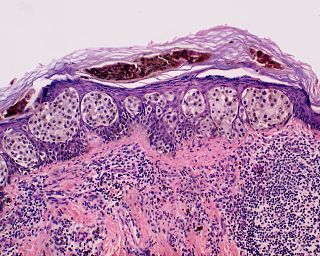
Pityriasis lichenoides et varioliformis acuta is a disease of the immune system. It is the more severe version of pityriasis lichenoides chronica. The disease is characterized by rashes and small lesions on the skin. The disease is more common in males and usually occurs in young adulthood, although it has been seen in every age group and every race. It is possible for the disease to go into remission for short periods of time or forever.

A skin condition, also known as cutaneous condition, is any medical condition that affects the integumentary system—the organ system that encloses the body and includes skin, nails, and related muscle and glands. The major function of this system is as a barrier against the external environment.
Granuloma faciale is an uncommon benign chronic skin disease of unknown origin characterized by single or multiple cutaneous nodules, usually occurring over the face. Occasionally, extrafacial involvement is noted, most often on sun-exposed areas.
Superficial spreading melanoma (SSM) is usually characterized as the most common form of cutaneous melanoma in Caucasians. The average age at diagnosis is in the fifth decade, and it tends to occur on sun-exposed skin, especially on the backs of males and lower limbs of females.

A blue nevus is a type of coloured mole, typically a single well-defined blue-black bump.
Acrodermatitis chronica atrophicans (ACA) is a skin rash indicative of the third or late stage of European Lyme borreliosis.
Erosive pustular dermatitis of the scalp presents with pustules, erosions, and crusts on the scalp of primarily older Caucasian females, and on biopsy, has a lymphoplasmacytic infiltrate with or without foreign body giant cells and pilosebaceous atrophy.
Papuloerythroderma of Ofuji is a rare disorder most commonly found in Japan, characterized by pruritic papules that spare the skinfolds, producing bands of uninvolved cutis, creating the so-called deck-chair sign. Frequently there is associated blood eosinophilia. Skin biopsies reveal a dense lymphohistiocytic infiltrate, eosinophils in the papillary dermis, and increased Langerhans cells. Systemic steroids are the treatment of choice and may result in long-term remissions.

Cutaneous small-vessel vasculitis (CSVV), also known as hypersensitivity vasculitis, cutaneous leukocytoclastic vasculitis, hypersensitivity angiitis, cutaneous leukocytoclastic angiitis, cutaneous necrotizing vasculitis and cutaneous necrotizing venulitis, is inflammation of small blood vessels, characterized by palpable purpura. It is the most common vasculitis seen in clinical practice.
Pseudomelanoma is a cutaneous condition in which melanotic skin lesions clinically resemble a superficial spreading melanoma at the site of a recent shave removal of a melanocytic nevus.