Dysembryoplastic neuroepithelial tumour

Last updated
Dysembryoplastic neuroepithelial tumour
DNET HE.jpg
DNET
Specialty Neuro-oncology, Neurosurgery

Dysembryoplastic neuroepithelial tumour (DNT, DNET) is a type of brain tumor. Most commonly found in the temporal lobe, DNTs have been classified as benign tumours. [1] These are glioneuronal tumours comprising both glial and neuron cells and often have ties to focal cortical dysplasia. [2]

Contents

Varying subclasses of DNTs have been presently identified, with dispute existing in the field on how to properly group these classes. [3] The identification of possible genetic markers to these tumours is currently underway. [4] With DNTs often causing epileptic seizures, surgical removal is a common treatment, providing high rates of success. [4]

Signs and symptoms

Seizures and epilepsy are the strongest ties to dysembryoplastic neuroepithelial tumours. [4] The most common symptom of DNTs are complex partial seizures. [2] Simple DNTs more frequently manifest generalized seizures. [2] In children, DNTs are considered to be the second leading cause of epilepsy. [3] A headache is another common symptom. [2] Diplopia may also be a result of a DNT. Other neurological impairments besides seizures are not common. [2]

Pathogenesis

Dysembryoplastic neuroepithelial tumours are largely glioneuronal tumours, meaning they are composed of both glial cells and neurons. [2]

Three subunits of DNTs have been commonly identified: [2]

There currently exists some debate over where to make the proper division for the subunits of DNTs. A fourth subunit is sometimes noted as a mixed subunit. This mixed subunit expresses the glial nodules and components of ganglioglioma. [1] Other findings suggest that DNTs require a reclassification to associate them with oligodendrogliomas, tumours that arise from solely glial cells. [3] These reports suggest that the neurons found within DNTs are much rarer than previously reported. For the neurons that are seen in the tumours, it is suggested that they had been trapped within the tumor upon formation, and are not a part of the tumour itself. [3]

Diagnosis

Dysembryoplastic neuroepithelial tumour, MRI FLAIR. DNET02.jpg
Dysembryoplastic neuroepithelial tumour, MRI FLAIR.

A dysembryoplastic neuroepithelial tumour is commonly diagnosed in patients who are experiencing seizures with magnetic resonance imaging (MRI), electroencephalogram (EEG). [4] A DNT is most commonly diagnosed in children who are experiencing seizures, and when given medication do not respond to them. When an MRI is taken there are lesions located in the temporal parietal region of the brain. [4]

Typical DNTs can be detected in an EEG scan when there are rapid repetitive spikes against a contrasted background. [4] EEG are predominantly localized with DNT location in the brain, however there are nonspecific cases in which the location of the tumour is abnormal and not localized. [4]

Classification

Dysembryoplastic neuroepithelial tumours are classified as a benign tumour, Grade I of the World Health Organization (WHO) classification of brain tumours. [1] This classification by WHO only covers the simple and complex subunits. Groups lacking glioneuronal elements were not considered to have fallen in the same group and have thusly not yet been classified. [1]

Complications in diagnosis

Dysembryoplastic neuroepithelial tumours are often described as a low grade tumour because about 1.2% people under the age of twenty are affected and about 0.2% over the age of twenty are affected by this tumour. [5] Since its prevalence is small among the population, it often goes misdiagnosed or even at times goes undiagnosed.[ citation needed ]

Treatment

The most common course of treatment of DNT is surgery. About 70-90% of surgery are successful in removing the tumour. [4] Since the tumour is most often benign, and does not impose immediate threat, aggressive treatments such as chemotherapy and radiation are not needed, and therefore patients especially children and young adults do not have to go through the side effects of these treatments. [5]

In order for the seizures to completely be stopped the tumour needs to be completely removed. For the tumor to be completely removed doctors need to perform resections consisting an anterior temporal lobectomy or amygdalo-hippocampectomy. [4] Alternatively, if the tumor is found at or near the surface of the brain, it can be removed without any other requirements. It has been found that if the tumour is removed by performing resections patients are then recognized as seizure free. On the other hand, if resections are not performed, and the tumour is not completely removed, then the patient is still at risk of experiencing the seizures. [4] In a study done by Bilginer et al., 2009, looking at patients whose tumour was not completely removed, and saw that they were still experiencing seizures, concluding that the incomplete resection as a being a failure. [4] This then causes the patient to undergo a second surgery and remove the tumour in which case causing a complete resection.[ citation needed ]

Outcomes

Recurrence of the tumour is highly unlikely if the patient undergoes a complete resection since the tumour is completely taken out. [5] Most of the tumours observed in patients are benign tumours, and once taken out do not cause neurological deficits. However, there have been incidents where the tumour was malignant. [5] There have been cases where the malignant tumour has made a reoccurrence, and this happens at the site of the residual tumour in which an incomplete resection has been done. [4] In this case, a second operation has to be done in order to completely remove the malignant tumour. In a study done with Daumas Duport and Varlet, 2003, they have found that there has been one case so far that the tumour has come back, however, in that particular case the patient underwent an incomplete resection, which led them to perform a second surgery in order to remove it completely. [4] This evidence shows that surgery and complete resections are one of the better approaches in treating dysembryoplastic neuroepithelial tumours.

Furthermore, a longer period of epilepsy, and patients older in age are less likely to have a full recovery and remain seizure free. This is the case because their body is not able to recover as quickly, as it would for a child who has had one seizure before. [5] Therefore, it is crucial to diagnose and perform the surgery early in order to make a full recovery.[ citation needed ]

Epidemiology

Children are much more prone to exhibit these dysembryoplastic neuroepithelial tumours than adults. [1] The mean age of onset of seizures for children with DNTs is 8.1 years old. [1] Few other neurological deficits are associated with DNTs, so that earlier detection of the tumour before seizure symptoms are rare. [2] DNTs are found in the temporal lobe in 84% of reported cases. [1] In children, DNTs account for 0.6% of diagnosed central nervous system tumours. [2] It has been found that males have a slightly higher risk of having these tumours. [2] Some familial accounts of DNTs have been documented, though the genetic ties have not yet been fully confirmed. [2]

History

Dysembryoplastic neuroepithelial tumours were usually found during investigation of patients who underwent multiple seizures. [2] The tumours were encountered when the patient required surgery to help with the epilepsy to help with the seizures. The term DNT was first introduced in 1988 by Daumas-Duport, terming it dysembryoplastic, suggesting a dysembryoplastic origin in early onset seizures, and neuroepithelial to allow the wide range of possible varieties of tumours to be put into the category. [2] In 2003 and 2007, DNT was made into further subsets of categories based upon the displayed elements within the tumour. [2]

See also

Related Research Articles

<span class="mw-page-title-main">Brain tumor</span> Neoplasm in the brain

A brain tumor occurs when abnormal cells form within the brain. There are two main types of tumors: malignant tumors and benign (non-cancerous) tumors. These can be further classified as primary tumors, which start within the brain, and secondary tumors, which most commonly have spread from tumors located outside the brain, known as brain metastasis tumors. All types of brain tumors may produce symptoms that vary depending on the size of the tumor and the part of the brain that is involved. Where symptoms exist, they may include headaches, seizures, problems with vision, vomiting and mental changes. Other symptoms may include difficulty walking, speaking, with sensations, or unconsciousness.

<span class="mw-page-title-main">Tuberous sclerosis</span> Genetic condition causing non-cancerous tumours

Tuberous sclerosis complex (TSC) is a rare multisystem autosomal dominant genetic disease that causes non-cancerous tumours to grow in the brain and on other vital organs such as the kidneys, heart, liver, eyes, lungs and skin. A combination of symptoms may include seizures, intellectual disability, developmental delay, behavioral problems, skin abnormalities, lung disease, and kidney disease.

<span class="mw-page-title-main">Focal cortical dysplasia</span> Medical condition

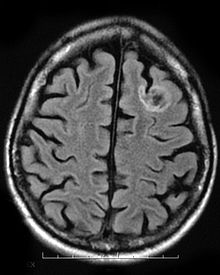
Focal cortical dysplasia (FCD) is a congenital abnormality of brain development where the neurons in an area of the brain failed to migrate in the proper formation in utero. Focal means that it is limited to a focal zone in any lobe. Focal cortical dysplasia is a common cause of intractable epilepsy in children and is a frequent cause of epilepsy in adults. There are three types of FCD with subtypes, including type 1a, 1b, 1c, 2a, 2b, 3a, 3b, 3c, and 3d, each with distinct histopathological features. All forms of focal cortical dysplasia lead to disorganization of the normal structure of the cerebral cortex :

<span class="mw-page-title-main">Neurofibromatosis type I</span> Type of neurofibromatosis disease

Neurofibromatosis type I (NF-1), or von Recklinghausen syndrome, is a complex multi-system human disorder caused by the mutation of Neurofibromin 1, a gene on chromosome 17 that is responsible for production of a protein (neurofibromin) which is needed for normal function in many human cell types. NF-1 causes tumors along the nervous system which can grow anywhere on the body. NF-1 is one of the most common genetic disorders and is not limited to any person's race or sex. NF-1 is an autosomal dominant disorder, which means that mutation or deletion of one copy of the NF-1 gene is sufficient for the development of NF-1, although presentation varies widely and is often different even between relatives affected by NF-1.

<span class="mw-page-title-main">Hamartoma</span> Tumour-like overgrowth due to a systemic genetic condition

A hamartoma is a mostly benign, local malformation of cells that resembles a neoplasm of local tissue but is usually due to an overgrowth of multiple aberrant cells, with a basis in a systemic genetic condition, rather than a growth descended from a single mutated cell (monoclonality), as would typically define a benign neoplasm/tumor. Despite this, many hamartomas are found to have clonal chromosomal aberrations that are acquired through somatic mutations, and on this basis the term hamartoma is sometimes considered synonymous with neoplasm. Hamartomas are by definition benign, slow-growing or self-limiting, though the underlying condition may still predispose the individual towards malignancies.

<span class="mw-page-title-main">Oligoastrocytoma</span> Medical condition

Oligoastrocytomas are a subset of brain tumors that present with an appearance of mixed glial cell origin, astrocytoma and oligodendroglioma. However, the term "Oligoastrocytoma" is now considered obsolete by the National Comprehensive Cancer Network stating "the term should no longer be used as such morphologically ambiguous tumors can be reliably resolved into astrocytomas and oligodendrogliomas with molecular testing."

<span class="mw-page-title-main">Gray matter heterotopia</span> Group of neurological disorders

Gray matter heterotopia is a neurological disorder caused by gray matter being located in an atypical location in the brain.

Neuroepithelial cells, or neuroectodermal cells, form the wall of the closed neural tube in early embryonic development. The neuroepithelial cells span the thickness of the tube's wall, connecting with the pial surface and with the ventricular or lumenal surface. They are joined at the lumen of the tube by junctional complexes, where they form a pseudostratified layer of epithelium called neuroepithelium.

<span class="mw-page-title-main">Choroid plexus papilloma</span> Medical condition

Choroid plexus papilloma, also known as papilloma of the choroid plexus, is a rare benign neuroepithelial intraventricular WHO grade I lesion found in the choroid plexus. It leads to increased cerebrospinal fluid production, thus causing increased intracranial pressure and hydrocephalus.

<span class="mw-page-title-main">Medulloepithelioma</span> Medical condition

Medulloepithelioma is a rare, primitive, fast-growing brain tumour thought to stem from cells of the embryonic medullary cavity. Tumours originating in the ciliary body of the eye are referred to as embryonal medulloepitheliomas, or diktyomas.

Neuro-oncology is the study of brain and spinal cord neoplasms, many of which are very dangerous and life-threatening. Among the malignant brain cancers, gliomas of the brainstem and pons, glioblastoma multiforme, and high-grade astrocytoma/oligodendroglioma are among the worst. In these cases, untreated survival usually amounts to only a few months, and survival with current radiation and chemotherapy treatments may extend that time from around a year to a year and a half, possibly two or more, depending on the patient's condition, immune function, treatments used, and the specific type of malignant brain neoplasm. Surgery may in some cases be curative, but, as a general rule, malignant brain cancers tend to regenerate and emerge from remission easily, especially highly malignant cases. In such cases, the goal is to excise as much of the mass and as much of the tumor margin as possible without endangering vital functions or other important cognitive abilities. The Journal of Neuro-Oncology is the longest continuously published journal in the field and serves as a leading reference to those practicing in the area of neuro-oncology.

<span class="mw-page-title-main">Fibrillary astrocytoma</span> Type of brain tumor

Fibrillary astrocytomas are a group of primary slow-growing brain tumors that typically occur in adults between the ages of 20 and 50.

<span class="mw-page-title-main">Tuber cinereum hamartoma</span> Human disease

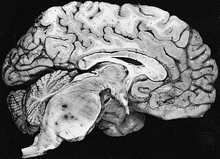
Tuber cinereum hamartoma is a benign tumor in which a disorganized collection of neurons and glia accumulate at the tuber cinereum of the hypothalamus on the floor of the third ventricle. It is a congenital malformation, included on the spectrum of gray matter heterotopias. Formation occurs during embryogenesis, typically between days 33 and 41 of gestation. Size of the tumor varies from one to three centimeters in diameter, with the mean being closer to the low end of this range. It is estimated to occur at a frequency of one in one million individuals.

<span class="mw-page-title-main">Subependymal giant cell astrocytoma</span> Medical condition

Subependymal giant cell astrocytoma is a low-grade astrocytic brain tumor (astrocytoma) that arises within the ventricles of the brain. It is most commonly associated with tuberous sclerosis complex (TSC). Although it is a low-grade tumor, its location can potentially obstruct the ventricles and lead to hydrocephalus.

<span class="mw-page-title-main">Astroblastoma</span> Medical condition

Astroblastoma is a rare glial tumor derived from the astroblast, a type of cell that closely resembles spongioblastoma and astrocytes. Astroblastoma cells are most likely found in the supratentorial region of the brain that houses the cerebrum, an area responsible for all voluntary movements in the body. It also occurs significantly in the frontal lobe, parietal lobe, and temporal lobe, areas where movement, language creation, memory perception, and environmental surroundings are expressed. These tumors can be present in major brain areas not associated with the main cerebral hemispheres, including the cerebellum, optic nerve, cauda equina, hypothalamus, and brain stem.

<span class="mw-page-title-main">Central neurocytoma</span> Medical condition

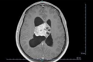
Central neurocytoma (CNC) is an extremely rare, ordinarily benign intraventricular brain tumour that typically forms from the neuronal cells of the septum pellucidum. The majority of central neurocytomas grow inwards into the ventricular system forming interventricular neurocytomas. This leads to two primary symptoms of CNCs, blurred vision and increased intracranial pressure. Treatment for a central neurocytoma typically involves surgical removal, with an approximate 1 in 5 chance of recurrence. Central neurocytomas are classified as a grade II tumor under the World Health Organization's classification of tumors of the nervous system.

<span class="mw-page-title-main">Ulegyria</span> Type of cortical scarring deep in the sulci

Ulegyria is a diagnosis used to describe a specific type of cortical scarring in the deep regions of the sulcus that leads to distortion of the gyri. Ulegyria is identified by its characteristic "mushroom-shaped" gyri, in which scarring causes shrinkage and atrophy in the deep sulcal regions while the surface gyri are spared. This condition is most often caused by hypoxic-ischemic brain injury in the perinatal period. The effects of ulegyria can range in severity, although it is most commonly associated with cerebral palsy, mental retardation and epilepsy. N.C. Bresler was the first to view ulegyria in 1899 and described this abnormal morphology in the brain as “mushroom-gyri." Although ulegyria was first identified in 1899, there is still limited information known or reported about the condition.

<span class="mw-page-title-main">James Rutka</span> Canadian neurosurgeon

James Rutka is a Canadian neurosurgeon from Toronto, Canada. Rutka served as RS McLaughlin Professor and Chair of the Department of Surgery in the Faculty of Medicine at the University of Toronto from 2011 – 2022. He subspecializes in pediatric neurosurgery at The Hospital for Sick Children (SickKids), and is a Senior Scientist in the Research Institute at SickKids. His main clinical interests include the neurosurgical treatment of children with brain tumours and epilepsy. His research interests lie in the molecular biology of human brain tumours – specifically in the determination of the mechanisms by which brain tumours grow and invade. He is the Director of the Arthur and Sonia Labatt Brain Tumour Research Centre at SickKids, and Editor-in-Chief of the Journal of Neurosurgery.

<span class="mw-page-title-main">Occipital epilepsy</span> Medical condition

Occipital epilepsy is a neurological disorder that arises from excessive neural activity in the occipital lobe of the brain that may or may not be symptomatic. Occipital lobe epilepsy is fairly rare, and may sometimes be misdiagnosed as migraine when symptomatic. Epileptic seizures are the result of synchronized neural activity that is excessive, and may stem from a failure of inhibitory neurons to regulate properly.

<span class="mw-page-title-main">Angiocentric glioma</span>

Angiocentric glioma (AG) refers to a rare neuroepithelial tumor when the superficial brain malignant cells enclose the brain vessels, commonly found in children and young adults. Initially identified in 2005 by Wang and his team from the University of Texas, AG was classified as Grade I by 2007 WHO Classification of Tumors of the Central Nervous System due to its benign clinical behavior, low proliferation index, and curative properties. AG primarily affects children and young adults at an average initial diagnosis age of 16 years old. Over 85% AG patients experience intractable seizures since childhood, especially partial epilepsy.

References

  1. 1 2 3 4 5 6 7 Thom M, Toma A, An S, Martinian L, Hadjivassiliou G, Ratilal B, et al. (October 2011). "One hundred and one dysembryoplastic neuroepithelial tumors: an adult epilepsy series with immunohistochemical, molecular genetic, and clinical correlations and a review of the literature". Journal of Neuropathology and Experimental Neurology. 70 (10): 859–878. doi: 10.1097/NEN.0b013e3182302475 . PMID   21937911.
  2. 1 2 3 4 5 6 7 8 9 10 11 12 13 14 15 16 17 18 Suh YL (November 2015). "Dysembryoplastic Neuroepithelial Tumors". Journal of Pathology and Translational Medicine. 49 (6): 438–449. doi:10.4132/jptm.2015.10.05. PMC   4696533 . PMID   26493957.
  3. 1 2 3 4 Komori T, Arai N (August 2013). "Dysembryoplastic neuroepithelial tumor, a pure glial tumor? Immunohistochemical and morphometric studies". Neuropathology. 33 (4): 459–468. doi: 10.1111/neup.12033 . PMID   23530928.
  4. 1 2 3 4 5 6 7 8 9 10 11 12 13 14 Chassoux F, Daumas-Duport C (December 2013). "Dysembryoplastic neuroepithelial tumors: where are we now?". Epilepsia. 54 (Suppl 9): 129–134. doi: 10.1111/epi.12457 . PMID   24328886.
  5. 1 2 3 4 5 Guduru H, Shen JK, Lokannavar HS (2012-01-01). "A rare case of dysembryoplastic neuroepithelial tumor". Journal of Clinical Imaging Science. 2 (1): 60. doi: 10.4103/2156-7514.102057 . PMC   3515966 . PMID   23230542.