Temporal lobe

Last updated

Temporal lobe
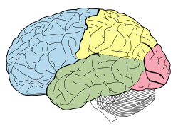
Lobes of the human brain (temporal lobe is shown in green)
Gray730.png
Section of brain showing upper surface of temporal lobe.
Details
Part of Cerebrum
Artery
Vein
Identifiers
Latin lobus temporalis
MeSH D013702
NeuroNames 125
NeuroLex ID birnlex_1160
TA98 A14.1.09.136
TA2 5488
FMA 61825
Anatomical terms of neuroanatomy

The temporal lobe is one of the four major lobes of the cerebral cortex in the brain of mammals. The temporal lobe is located beneath the lateral fissure on both cerebral hemispheres of the mammalian brain. [3]

Contents

The temporal lobe is involved in processing sensory input into derived meanings for the appropriate retention of visual memory, language comprehension, and emotion association. [4] :21Temporal refers to the head's temples.

Structure

The temporal lobe consists of structures that are vital for declarative or long-term memory. Declarative (denotative) or explicit memory is conscious memory divided into semantic memory (facts) and episodic memory (events). [4] :194 Medial temporal lobe structures that are critical for long-term memory include the hippocampus, along with the surrounding hippocampal region consisting of the perirhinal, parahippocampal, and entorhinal neocortical regions. [4] :196 The hippocampus is critical for memory formation, and the surrounding medial temporal cortex is currently theorized to be critical for memory storage. [4] :21 The prefrontal and visual cortices are also involved in explicit memory. [4] :21

Research has shown that lesions in the hippocampus of monkeys results in limited impairment of function, whereas extensive lesions that include the hippocampus and the medial temporal cortex result in severe impairment. [5]

Function

Visual memories

The temporal lobe communicates with the hippocampus and plays a key role in the formation of explicit long-term memory modulated by the amygdala. [4] :349

Processing sensory input

Auditory
Adjacent areas in the superior, posterior, and lateral parts of the temporal lobes are involved in high-level auditory processing. The temporal lobe is involved in primary auditory perception, such as hearing, and holds the primary auditory cortex. [6] The primary auditory cortex receives sensory information from the ears and secondary areas process the information into meaningful units such as speech and words. [6] The superior temporal gyrus includes an area (within the lateral fissure) where auditory signals from the cochlea first reach the cerebral cortex and are processed by the primary auditory cortex in the left temporal lobe.[ citation needed ]
Visual
The areas associated with vision in the temporal lobe interpret the meaning of visual stimuli[ clarification needed ] and establish object recognition. [7] The ventral part of the temporal cortices appears to be involved in high-level visual processing of complex stimuli such as faces (fusiform gyrus) [8] and scenes (parahippocampal gyrus). [9] Anterior parts of this ventral stream for visual processing are involved in object perception and recognition. [6]
Animation showing the position of the human left temporal lobe Temporal lobe animation.gif
Animation showing the position of the human left temporal lobe

Language recognition

In humans, temporal lobe regions are critical for accessing the semantic meaning of spoken words, printed words, and visual objects [10] . Wernicke's area, which spans the region between temporal and parietal lobes of the dominant cerebral hemisphere (the left, in the majority of cases), plays a key role (in tandem with Broca's area in the frontal lobe) in language comprehension, [11] whether spoken language or signed language. FMRI imaging shows these portions of the brain are activated by signed or spoken languages. [12] [13] These areas of the brain are active in children's language acquisition [14] whether accessed via hearing a spoken language, watching a signed language, or via hand-over-hand tactile versions of a signed language. [15]

The functions of the left temporal lobe are not limited to low-level perception but extend to comprehension, naming, and verbal memory. [16]

New memories

The medial temporal lobes (near the sagittal plane) are thought to be involved in encoding declarative long term memory. [4] :194–199 The medial temporal lobes include the hippocampi, which are essential for memory storage, therefore damage to this area can result in impairment in new memory formation leading to permanent or temporary anterograde amnesia. [4] :194–199

Clinical significance

Unilateral temporal lesion

Dominant hemisphere

Non-dominant hemisphere

Bitemporal lesions (additional features)

Damage

Individuals who suffer from medial temporal lobe damage have a difficult time recalling visual stimuli. This neurotransmission deficit is not due to lacking perception of visual stimuli, but rather to the inability to interpret what is perceived. [17] The most common symptom of inferior temporal lobe damage is visual agnosia, which involves impairment in the identification of familiar objects. Another less common type of inferior temporal lobe damage is prosopagnosia which is an impairment in the recognition of faces and distinction of unique individual facial features. [18]

Damage specifically to the anterior portion of the left temporal lobe can cause savant syndrome. [19]

Disorders

Pick's disease, also known as frontotemporal amnesia, is caused by atrophy of the frontotemporal lobe. [20] Emotional symptoms include mood changes, which the patient may be unaware of, including poor attention span and aggressive behavior towards themselves or others. Language symptoms include loss of speech, inability to read or write, loss of vocabulary and overall degeneration of motor ability. [21]

Temporal lobe epilepsy is a chronic neurological condition characterized by recurrent seizures; symptoms include a variety of sensory (visual, auditory, olfactory, and gustation) hallucinations, as well as an inability to process semantic and episodic memories. [22]

Schizophrenia is a severe psychotic disorder characterized by severe disorientation. Its most explicit symptom is the perception of external voices in the form of auditory hallucinations. The cause of such hallucinations has been attributed to deficits in the left temporal lobe, specifically within the primary auditory cortex. [23] [24] Decreased gray matter, among other cellular deficits, contribute to spontaneous neural activity that affects the primary auditory cortex as if it were experiencing acoustic auditory input. The misrepresentation of speech in the auditory cortex results in the perception of external voices in the form of auditory hallucinations in schizophrenic patients. [25] Structural and functional MRI techniques have accounted for this neural activity by testing affected and non-affected individuals with external auditory stimuli. [23]

See also

Related Research Articles

<span class="mw-page-title-main">Entorhinal cortex</span> Area of the temporal lobe of the brain

The entorhinal cortex (EC) is an area of the brain's allocortex, located in the medial temporal lobe, whose functions include being a widespread network hub for memory, navigation, and the perception of time. The EC is the main interface between the hippocampus and neocortex. The EC-hippocampus system plays an important role in declarative (autobiographical/episodic/semantic) memories and in particular spatial memories including memory formation, memory consolidation, and memory optimization in sleep. The EC is also responsible for the pre-processing (familiarity) of the input signals in the reflex nictitating membrane response of classical trace conditioning; the association of impulses from the eye and the ear occurs in the entorhinal cortex.

<span class="mw-page-title-main">Parietal lobe</span> Part of the brain responsible for sensory input and some language processing

The parietal lobe is one of the four major lobes of the cerebral cortex in the brain of mammals. The parietal lobe is positioned above the temporal lobe and behind the frontal lobe and central sulcus.

<span class="mw-page-title-main">Fusiform gyrus</span> Gyrus of the temporal and occipital lobes of the brain

The fusiform gyrus, also known as the lateral occipitotemporal gyrus,is part of the temporal lobe and occipital lobe in Brodmann area 37. The fusiform gyrus is located between the lingual gyrus and parahippocampal gyrus above, and the inferior temporal gyrus below. Though the functionality of the fusiform gyrus is not fully understood, it has been linked with various neural pathways related to recognition. Additionally, it has been linked to various neurological phenomena such as synesthesia, dyslexia, and prosopagnosia.

<span class="mw-page-title-main">Auditory cortex</span> Part of the temporal lobe of the brain

The auditory cortex is the part of the temporal lobe that processes auditory information in humans and many other vertebrates. It is a part of the auditory system, performing basic and higher functions in hearing, such as possible relations to language switching. It is located bilaterally, roughly at the upper sides of the temporal lobes – in humans, curving down and onto the medial surface, on the superior temporal plane, within the lateral sulcus and comprising parts of the transverse temporal gyri, and the superior temporal gyrus, including the planum polare and planum temporale.

Visual processing is a term that is used to refer to the brain's ability to use and interpret visual information from the world. The process of converting light energy into a meaningful image is a complex process that is facilitated by numerous brain structures and higher level cognitive processes. On an anatomical level, light energy first enters the eye through the cornea, where the light is bent. After passing through the cornea, light passes through the pupil and then lens of the eye, where it is bent to a greater degree and focused upon the retina. The retina is where a group of light-sensing cells, called photoreceptors are located. There are two types of photoreceptors: rods and cones. Rods are sensitive to dim light and cones are better able to transduce bright light. Photoreceptors connect to bipolar cells, which induce action potentials in retinal ganglion cells. These retinal ganglion cells form a bundle at the optic disc, which is a part of the optic nerve. The two optic nerves from each eye meet at the optic chiasm, where nerve fibers from each nasal retina cross which results in the right half of each eye's visual field being represented in the left hemisphere and the left half of each eye's visual fields being represented in the right hemisphere. The optic tract then diverges into two visual pathways, the geniculostriate pathway and the tectopulvinar pathway, which send visual information to the visual cortex of the occipital lobe for higher level processing.

The entorhinal cortex (EC) is a major part of the hippocampal formation of the brain, and is reciprocally connected with the hippocampus.

<span class="mw-page-title-main">Language processing in the brain</span> How humans use words to communicate

In psycholinguistics, language processing refers to the way humans use words to communicate ideas and feelings, and how such communications are processed and understood. Language processing is considered to be a uniquely human ability that is not produced with the same grammatical understanding or systematicity in even human's closest primate relatives.

In neurology, semantic dementia (SD), also known as semantic variant primary progressive aphasia (svPPA), is a progressive neurodegenerative disorder characterized by loss of semantic memory in both the verbal and non-verbal domains. However, the most common presenting symptoms are in the verbal domain. Semantic dementia is a disorder of semantic memory that causes patients to lose the ability to match words or images to their meanings. However, it is fairly rare for patients with semantic dementia to develop category specific impairments, though there have been documented cases of it occurring. Typically, a more generalized semantic impairment results from dimmed semantic representations in the brain.

<span class="mw-page-title-main">Cingulum (brain)</span> Nerve tract from the cingulate gyrus to the entorhinal cortex in the brain

In neuroanatomy, the cingulum or cingulum bundle is an association tract, a nerve tract that projects from the cingulate gyrus to the entorhinal cortex in the brain, allowing for communication between components of the limbic system. It forms the white matter core of the cingulate gyrus, following it from the subcallosal gyrus of the frontal lobe beneath the rostrum of corpus callosum to the parahippocampal gyrus and uncus of the temporal lobe.

<span class="mw-page-title-main">Parahippocampal gyrus</span> Grey matter region surrounding the hippocampus

The parahippocampal gyrus is a grey matter cortical region of the brain that surrounds the hippocampus and is part of the limbic system. The region plays an important role in memory encoding and retrieval. It has been involved in some cases of hippocampal sclerosis. Asymmetry has been observed in schizophrenia.

<span class="mw-page-title-main">Lobes of the brain</span> Parts of the cerebrum

The lobes of the brain are the major identifiable zones of the human cerebral cortex, and they comprise the surface of each hemisphere of the cerebrum. The two hemispheres are roughly symmetrical in structure, and are connected by the corpus callosum. They traditionally have been divided into four lobes, but are today considered as having six lobes each. The lobes are large areas that are anatomically distinguishable, and are also functionally distinct to some degree. Each lobe of the brain has numerous ridges, or gyri, and furrows, the sulci that constitute further subzones of the cortex. The expression "lobes of the brain" usually refers only to those of the cerebrum, not to the distinct areas of the cerebellum.

The two-streams hypothesis is a model of the neural processing of vision as well as hearing. The hypothesis, given its initial characterisation in a paper by David Milner and Melvyn A. Goodale in 1992, argues that humans possess two distinct visual systems. Recently there seems to be evidence of two distinct auditory systems as well. As visual information exits the occipital lobe, and as sound leaves the phonological network, it follows two main pathways, or "streams". The ventral stream leads to the temporal lobe, which is involved with object and visual identification and recognition. The dorsal stream leads to the parietal lobe, which is involved with processing the object's spatial location relative to the viewer and with speech repetition.

<span class="mw-page-title-main">Inferior temporal gyrus</span> One of three gyri of the temporal lobe of the brain

The inferior temporal gyrus is one of three gyri of the temporal lobe and is located below the middle temporal gyrus, connected behind with the inferior occipital gyrus; it also extends around the infero-lateral border on to the inferior surface of the temporal lobe, where it is limited by the inferior sulcus. This region is one of the higher levels of the ventral stream of visual processing, associated with the representation of objects, places, faces, and colors. It may also be involved in face perception, and in the recognition of numbers and words.

<span class="mw-page-title-main">Lingual gyrus</span> Gyrus of the occipital lobe of the brain

The lingual gyrus, also known as the medialoccipitotemporal gyrus, is a brain structure that is linked to processing vision, especially related to letters. It is thought to also play a role in analysis of logical conditions and encoding visual memories. It is named after its shape, which is somewhat similar to a tongue. Contrary to the name, the region has little to do with speech.

The perirhinal cortex is a cortical region in the medial temporal lobe that is made up of Brodmann areas 35 and 36. It receives highly processed sensory information from all sensory regions, and is generally accepted to be an important region for memory. It is bordered caudally by postrhinal cortex or parahippocampal cortex and ventrally and medially by entorhinal cortex.

The neuroanatomy of memory encompasses a wide variety of anatomical structures in the brain.

Recognition memory, a subcategory of explicit memory, is the ability to recognize previously encountered events, objects, or people. When the previously experienced event is reexperienced, this environmental content is matched to stored memory representations, eliciting matching signals. As first established by psychology experiments in the 1970s, recognition memory for pictures is quite remarkable: humans can remember thousands of images at high accuracy after seeing each only once and only for a few seconds.

<span class="mw-page-title-main">Superior temporal sulcus</span> Part of the brains temporal lobe

In the human brain, the superior temporal sulcus (STS) is the sulcus separating the superior temporal gyrus from the middle temporal gyrus in the temporal lobe of the brain. A sulcus is a deep groove that curves into the largest part of the brain, the cerebrum, and a gyrus is a ridge that curves outward of the cerebrum.

Visual object recognition refers to the ability to identify the objects in view based on visual input. One important signature of visual object recognition is "object invariance", or the ability to identify objects across changes in the detailed context in which objects are viewed, including changes in illumination, object pose, and background context.

<span class="mw-page-title-main">Auditosensory cortex</span>

The auditosensory cortex is the part of the auditory system that is associated with the sense of hearing in humans. It occupies the bilateral primary auditory cortex in the temporal lobe of the mammalian brain. The term is used to describe Brodmann areas 41 and 42 together with the transverse temporal gyrus. The auditosensory cortex takes part in the reception and processing of auditory nerve impulses, which passes sound information from the thalamus to the brain. Abnormalities in this region are responsible for many disorders in auditory abilities, such as congenital deafness, true cortical deafness, primary progressive aphasia and auditory hallucination.

References

  1. 1 2 3 Starr PA, Barbaro NM, Larson PS (30 November 2008). Neurosurgical Operative Atlas: Functional Neurosurgery. Thieme. pp. 16, 26. ISBN   978-1-58890-399-0.
  2. Sekhar LN, de Oliveira E (1999). Cranial Microsurgery: Approaches and Techniques. Thieme. p. 432. ISBN   978-0-86577-698-2.
  3. "Temporal Lobe". Langbrain. Rice University. Retrieved 2 January 2011.
  4. 1 2 3 4 5 6 7 8 Smith EE, Kosslyn SM (2007). Cognitive Psychology: Mind and Brain. New Jersey: Prentice Hall. pp. 21, 194–199, 349. ISBN   978-0-13-182508-6.
  5. Squire LR, Stark CE, Clark RE (2004). "The medial temporal lobe". Annual Review of Neuroscience. 27: 279–306. doi:10.1146/annurev.neuro.27.070203.144130. PMID   15217334.
  6. 1 2 3 Schacter DL, Gilbert DT, Wegner DM (2010). Psychology (2nd ed.). New York: Worth Publishers. ISBN   978-1-4292-3719-2.[ page needed ]
  7. Okamoto N, Seiyama A, Hori S, Takahashi S (2024-05-03). "Role of the left posterior middle temporal gyrus in shape recognition and its reconstruction during drawing: A study combining transcranial magnetic stimulation and functional near infrared spectroscopy". PLOS ONE. 19 (5): e0302375. Bibcode:2024PLoSO..1902375O. doi: 10.1371/journal.pone.0302375 . PMC   11068196 . PMID   38701103.
  8. Volfart A, Jonas J, Maillard L, Colnat-Coulbois S, Rossion B (April 2020). Freiwald WA (ed.). "Neurophysiological evidence for crossmodal (face-name) person-identity representation in the human left ventral temporal cortex". PLOS Biology. 18 (4): e3000659. doi: 10.1371/journal.pbio.3000659 . PMC   7159237 . PMID   32243450.
  9. Bastin J, Committeri G, Kahane P, Galati G, Minotti L, Lachaux JP, et al. (June 2013). "Timing of posterior parahippocampal gyrus activity reveals multiple scene processing stages". Human Brain Mapping. 34 (6): 1357–1370. doi:10.1002/hbm.21515. PMC   6870532 . PMID   22287281.
  10. Visser M, Jefferies E, Lambon Ralph MA (June 2010). "Semantic Processing in the Anterior Temporal Lobes: A Meta-analysis of the Functional Neuroimaging Literature". Journal of Cognitive Neuroscience. 22 (6): 1083–1094. doi:10.1162/jocn.2009.21309. PMID   19583477.
  11. Hickok G, Poeppel D (May 2007). "The cortical organization of speech processing". Nature Reviews. Neuroscience. 8 (5): 393–402. doi:10.1038/nrn2113. PMID   17431404. S2CID   6199399.
  12. Richardson MW. "Does the Brain Process Sign Language and Spoken Language Differently?". www.brainfacts.org. Retrieved 2020-12-14.
  13. Campbell R, MacSweeney M, Waters D (29 June 2007). "Sign language and the brain: a review". Journal of Deaf Studies and Deaf Education. 13 (1): 3–20. doi: 10.1093/deafed/enm035 . PMID   17602162.
  14. "Language Learning Through the Eye and Ear Webcast". clerccenter.gallaudet.edu. Archived from the original on 2020-12-05. Retrieved 2020-12-16.
  15. Humphries T, Kushalnagar P, Mathur G, Napoli DJ, Padden C, Rathmann C, et al. (April 2012). "Language acquisition for deaf children: Reducing the harms of zero tolerance to the use of alternative approaches". Harm Reduction Journal. 9: 16. doi: 10.1186/1477-7517-9-16 . PMC   3384464 . PMID   22472091.
  16. Mitjana LR (6 September 2019). "Lóbulo temporal: anatomía, funciones y características" [Temporal lobe: anatomy, functions and characteristics]. MedSalud (in Spanish).
  17. Pertzov Y, Miller TD, Gorgoraptis N, Caine D, Schott JM, Butler C, et al. (August 2013). "Binding deficits in memory following medial temporal lobe damage in patients with voltage-gated potassium channel complex antibody-associated limbic encephalitis". Brain: A Journal of Neurology. 136 (Pt 8): 2474–2485. doi:10.1093/brain/awt129. ISSN   1460-2156. PMC   3722347 . PMID   23757763.
  18. Mizuno T, Takeda K (November 2009). "[The symptomatology of frontal and temporal lobe damages]". Brain and Nerve = Shinkei Kenkyu No Shinpo (in Japanese). 61 (11): 1209–18. PMID   19938677.
  19. Treffert DA (May 2009). "The savant syndrome: an extraordinary condition. A synopsis: past, present, future". Philosophical Transactions of the Royal Society of London. Series B, Biological Sciences. 364 (1522): 1351–1357. doi:10.1098/rstb.2008.0326. PMC   2677584 . PMID   19528017.
  20. Takeda N, Kishimoto Y, Yokota O (2012). "Pick's Disease". Neurodegenerative Diseases. Advances in Experimental Medicine and Biology. Vol. 724. pp. 300–316. doi:10.1007/978-1-4614-0653-2_23. ISBN   978-1-4614-0652-5. PMID   22411252.
  21. Yokota O, Tsuchiya K, Arai T, Yagishita S, Matsubara O, Mochizuki A, et al. (April 2009). "Clinicopathological characterization of Pick's disease versus frontotemporal lobar degeneration with ubiquitin/TDP-43-positive inclusions". Acta Neuropathologica. 117 (4): 429–444. doi:10.1007/s00401-009-0493-4. PMID   19194716. S2CID   23749655.
  22. Lah S, Smith ML (January 2014). "Semantic and episodic memory in children with temporal lobe epilepsy: do they relate to literacy skills?". Neuropsychology. 28 (1): 113–22. doi:10.1037/neu0000029. PMID   24245928.
  23. 1 2 Hugdahl K, Løberg EM, Nygård M (May 2009). "Left temporal lobe structural and functional abnormality underlying auditory hallucinations in schizophrenia". Frontiers in Neuroscience. 3 (1): 34–45. doi: 10.3389/neuro.01.001.2009 . PMC   2695389 . PMID   19753095.
  24. Hugdahl K, Løberg EM, Nygård M (May 2009). "Left temporal lobe structural and functional abnormality underlying auditory hallucinations in schizophrenia". Frontiers in Neuroscience. 3 (1): 34–45. doi: 10.3389/neuro.01.001.2009 . PMC   2695389 . PMID   19753095.
  25. Ikuta T, DeRosse P, Argyelan M, Karlsgodt KH, Kingsley PB, Szeszko PR, et al. (December 2015). "Subcortical modulation in auditory processing and auditory hallucinations". Behavioural Brain Research. 295: 78–81. doi:10.1016/j.bbr.2015.08.009. PMC   4641005 . PMID   26275927.