Additional images
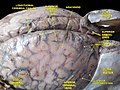 Meninges and superficial cerebral veins. Deep dissection. Superior view.
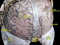 Meninges and superficial cerebral veins. Deep dissection. Superior view.
| Inferior anastomotic vein | |
|---|---|
| Details | |
| Identifiers | |
| Latin | vena anastomotica inferior |
| TA98 | A12.3.06.010 |
| TA2 | 4910 |
| FMA | 51239 |
| Anatomical terminology | |
The inferior anastomotic vein (also known as the vein of Labbe) is one of several superficial cerebral veins.[ citation needed ] It is a large, [1] highly variable[ citation needed ] vein extending across the lateral hemispheric surface of the temporal lobe to form an anastomosis between the superficial middle cerebral vein and transverse sinus, opening into either at either end. [1] It drains adjacent cortical regions, gathering tributaries from minor veins of the temporal lobe.[ citation needed ]
It was named after the 19th century French surgeon Charles Labbé (1851–1889), the nephew of the surgeon and politician Léon Labbé (1832–1916).


| | This cardiovascular system article is a stub. You can help Wikipedia by expanding it. |