Veins are blood vessels in the circulatory system of humans and most other animals that carry blood towards the heart. Most veins carry deoxygenated blood from the tissues back to the heart; exceptions are those of the pulmonary and fetal circulations which carry oxygenated blood to the heart. In the systemic circulation, arteries carry oxygenated blood away from the heart, and veins return deoxygenated blood to the heart, in the deep veins.

Pia mater, often referred to as simply the pia, is the delicate innermost layer of the meninges, the membranes surrounding the brain and spinal cord. Pia mater is medieval Latin meaning "tender mother". The other two meningeal membranes are the dura mater and the arachnoid mater. Both the pia and arachnoid mater are derivatives of the neural crest while the dura is derived from embryonic mesoderm. The pia mater is a thin fibrous tissue that is permeable to water and small solutes. The pia mater allows blood vessels to pass through and nourish the brain. The perivascular space between blood vessels and pia mater is proposed to be part of a pseudolymphatic system for the brain. When the pia mater becomes irritated and inflamed the result is meningitis.

The internal carotid artery is an artery in the neck which supplies the anterior and middle cerebral circulation.
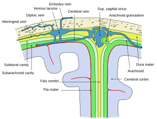
Arachnoid granulations are small outpouchings of the arachnoid mater and subarachnoid space into the dural venous sinuses of the brain. The granulations are thought to mediate the draining of cerebrospinal fluid (CSF) from the subarachnoid space into the venous system.

The dura mater, is the outermost of the three meningeal membranes. The dura mater has two layers, an outer periosteal layer closely adhered to the neurocranium, and an inner meningeal layer known as the dural border cell layer. The two dural layers are for the most part fused together forming a thick fibrous tissue membrane that covers the brain and the vertebrae of the spinal column. But the layers are separated at the dural venous sinuses to allow blood to drain from the brain. The dura covers the arachnoid mater and the pia mater the other two meninges in protecting the central nervous system.

The great cerebral vein is one of the large blood vessels in the skull draining the cerebrum of the brain. It is also known as the vein of Galen, named for its discoverer, the Greek physician Galen.

Cerebral circulation is the movement of blood through a network of cerebral arteries and veins supplying the brain. The rate of cerebral blood flow in an adult human is typically 750 milliliters per minute, or about 15% of cardiac output. Arteries deliver oxygenated blood, glucose and other nutrients to the brain. Veins carry "used or spent" blood back to the heart, to remove carbon dioxide, lactic acid, and other metabolic products. The neurovascular unit regulates cerebral blood flow so that activated neurons can be supplied with energy in the right amount and at the right time. Because the brain would quickly suffer damage from any stoppage in blood supply, the cerebral circulatory system has safeguards including autoregulation of the blood vessels. The failure of these safeguards may result in a stroke. The volume of blood in circulation is called the cerebral blood flow. Sudden intense accelerations change the gravitational forces perceived by bodies and can severely impair cerebral circulation and normal functions to the point of becoming serious life-threatening conditions.

Intracranial hemorrhage (ICH), also known as intracranial bleed, is bleeding within the skull. Subtypes are intracerebral bleeds, subarachnoid bleeds, epidural bleeds, and subdural bleeds.

In anatomy, the epidural space is the potential space between the dura mater and vertebrae (spine).

Epidural hematoma is when bleeding occurs between the tough outer membrane covering the brain and the skull. When this condition occurs in the spinal canal, it is known as a spinal epidural hematoma.

The falx cerebri is a large, crescent-shaped fold of dura mater that descends vertically into the longitudinal fissure to separate the cerebral hemispheres. It supports the dural sinuses that provide venous and CSF drainage from the brain. It is attached to the crista galli anteriorly, and blends with the tentorium cerebelli posteriorly.

The cavernous sinus within the human head is one of the dural venous sinuses creating a cavity called the lateral sellar compartment bordered by the temporal bone of the skull and the sphenoid bone, lateral to the sella turcica.
The straight sinus, also known as tentorial sinus or the sinus rectus, is an area within the skull beneath the brain. It receives blood from the inferior sagittal sinus and the great cerebral vein, and drains into the confluence of sinuses.
The superior sagittal sinus, within the human head, is an unpaired dural venous sinus lying along the attached margin of the falx cerebri. It allows blood to drain from the lateral aspects of anterior cerebral hemispheres to the confluence of sinuses. Cerebrospinal fluid drains through arachnoid granulations into the superior sagittal sinus and is returned to venous circulation.
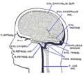
The inferior sagittal sinus, within the human head, is an area beneath the brain which allows blood to drain outwards posteriorly from the center of the head. It drains to the straight sinus, which connects to the transverse sinuses. See diagram : labeled in the brain as "SIN. SAGITTALIS INF.".
The sigmoid sinuses, also known as the pars sigmoid, are paired dural venous sinuses within the skull that receive blood from posterior transverse sinuses.
The transverse sinuses, within the human head, are two areas beneath the brain which allow blood to drain from the back of the head. They run laterally in a groove along the interior surface of the occipital bone. They drain from the confluence of sinuses to the sigmoid sinuses, which ultimately connect to the internal jugular vein. See diagram : labeled under the brain as "SIN. TRANS.".

Anterior spinal veins are veins that receive blood from the anterior spinal cord.
Sinus pericranii (SP) is a rare disorder characterized by a congenital epicranial venous malformation of the scalp. Treatment of this condition has mainly been recommended for aesthetic reasons and prevention of bleeding.
The empty delta sign is a radiologic sign seen on brain imaging which is associated with cerebral venous sinus thrombosis. It is usually seen on magnetic resonance imaging (MRI) or computed tomography (CT) scans with contrast. It is seen as dural wall enhancement in the absence of intra-sinus enhancement. This is due to the presence of a blood clot in the dural venous sinuses. The dural venous sinuses drain blood from the brain to the internal jugular veins, which in turn drains blood to the heart. It has been proposed that the empty delta sign occurs in dural venous thromboses due to contrast material filling the dural venous collateral circulation immediately surrounding the dura whilst being unable to fill the intra-dural sinus space due to the presence of a blood clot. The superior sagittal sinus is most commonly affected, but the radiologic sign may also be seen in the transverse sinuses.