The transverse sinuses (left and right lateral sinuses), within the human head, are two areas beneath the brain which allow blood to drain from the back of the head. They run laterally in a groove along the interior surface of the occipital bone. They drain from the confluence of sinuses (by the internal occipital protuberance) to the sigmoid sinuses, which ultimately connect to the internal jugular vein. See diagram (at right): labeled under the brain as "SIN. TRANS." (for Latin: sinus transversus).
Each transverse sinus passes lateral and forward, describing a slight curve with its convexity upward, to the base of the petrous portion of the temporal bone, and lies, in this part of its course, in the attached margin of the tentorium cerebelli; it then leaves the tentorium and curves downward and medialward (an area sometimes referred to as the sigmoid sinus) to reach the jugular foramen, where it ends in the internal jugular vein.
The transverse sinuses are frequently of unequal size, with the one formed by the superior sagittal sinus being the larger; they increase in size as they proceed, from back to center.
On transverse section, the horizontal portion exhibits a prismatic form, the curved portion has a semicylindrical form.
The petrosquamous sinus, when present, runs backward along the junction of the squama and petrous portion of the temporal, and opens into the transverse sinus.
Additional images
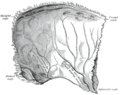
Left parietal bone. Inner surface.
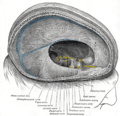
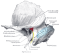
Dura mater and its processes exposed by removing part of the right half of the skull, and the brain
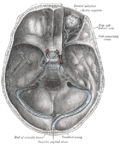
The sinuses at the base of the skull
Horizontal section through left ear; upper half of section
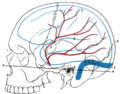
Relations of the brain and middle meningeal artery to the surface of the skull
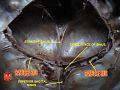
Left temporal bone showing surface markings for the tympanic antrum (red), transverse sinus (blue), and facial nerve (yellow)
This page is based on this Wikipedia article Text is available under the CC BY-SA 4.0 license; additional terms may apply. Images, videos and audio are available under their respective licenses.