Additional images
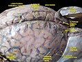 Meninges and superficial cerebral veins. Deep dissection. Superior view.
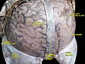 Meninges and superficial cerebral veins. Deep dissection. Superior view.
| Superior anastomotic vein | |
|---|---|
| Details | |
| Identifiers | |
| Latin | vena anastomotica superior |
| TA98 | A12.3.06.012 |
| TA2 | 4909 |
| FMA | 51238 |
| Anatomical terminology | |
The superior anastomotic vein, also known as the vein of Trolard, is a superficial cerebral vein grouped with the superior cerebral veins. The vein was eponymously named after the 18th century anatomist Jean Baptiste Paulin Trolard. The vein anastomoses with the middle cerebral vein and the superior sagittal sinus.


| | This cardiovascular system article is a stub. You can help Wikipedia by expanding it. |

The great saphenous vein (GSV) or long saphenous vein is a large, subcutaneous, superficial vein of the leg. It is the longest vein in the body, running along the length of the lower limb, returning blood from the foot, leg and thigh to the deep femoral vein at the femoral triangle.

The internal carotid artery is an artery in the neck which supplies the anterior circulation of the brain.

In neuroanatomy, dura mater is a thick membrane made of dense irregular connective tissue that surrounds the brain and spinal cord. It is the outermost of the three layers of membrane called the meninges that protect the central nervous system. The other two meningeal layers are the arachnoid mater and the pia mater. It envelops the arachnoid mater, which is responsible for keeping in the cerebrospinal fluid. It is derived primarily from the neural crest cell population, with postnatal contributions of the paraxial mesoderm.

In neuroanatomy, the optic tract is a part of the visual system in the brain. It is a continuation of the optic nerve that relays information from the optic chiasm to the ipsilateral lateral geniculate nucleus (LGN), pretectal nuclei, and superior colliculus.

The longitudinal fissure is the deep groove that separates the two cerebral hemispheres of the vertebrate brain. Lying within it is a continuation of the dura mater called the falx cerebri. The inner surfaces of the two hemispheres are convoluted by gyri and sulci just as is the outer surface of the brain.

In neuroanatomy, the corona radiata is a white matter sheet that continues inferiorly as the internal capsule and superiorly as the centrum semiovale. This sheet of both ascending and descending axons carries most of the neural traffic from and to the cerebral cortex. The corona radiata is associated with the corticopontine tract, the corticobulbar tract, and the corticospinal tract.

The external capsule is a series of white matter fiber tracts in the brain. These fibers run between the most lateral segment of the lentiform nucleus and the claustrum.

The subarachnoid cisterns are spaces formed by openings in the subarachnoid space, an anatomic space in the meninges of the brain. The space is situated between the two meninges, the arachnoid mater and the pia mater. These cisterns are filled with cerebrospinal fluid (CSF).

The arachnoid mater is one of the three meninges, the protective membranes that cover the brain and spinal cord. It is so named because of its resemblance to a spider web. The arachnoid mater is a derivative of the neural crest mesoectoderm in the embryo.

The internal iliac artery is the main artery of the pelvis.

The superior sagittal sinus, within the human head, is an unpaired area along the attached margin of the falx cerebri. It allows blood to drain from the lateral aspects of anterior cerebral hemispheres to the confluence of sinuses. Cerebrospinal fluid drains through arachnoid granulations into the superior sagittal sinus and is returned to venous circulation.

The inferior cerebral veins are veins that drain the undersurface of the cerebral hemispheres and empty into the cavernous and transverse sinuses.

The superior cerebral veins are several cerebral veins that drain the superolateral and superomedial surfaces of the cerebral hemispheres into the superior sagittal sinus. There are 8-12 cerebral veins. They are predominantly found in the sulci between the gyri, but can also be found running across the gyri.

In human anatomy, the cerebral veins are blood vessels in the cerebral circulation which drain blood from the cerebrum of the human brain. They are divisible into external and internal groups according to the outer or inner parts of the hemispheres they drain into.
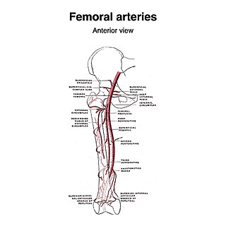
The superficial iliac circumflex artery, the smallest of the cutaneous branches of the femoral artery, arises close to the superficial epigastric artery, and, piercing the fascia lata, runs lateralward, parallel with the inguinal ligament, as far as the crest of the ilium.

The external pudendal veins are veins of the pelvis which drain into the great saphenous vein.

The following outline is provided as an overview of and topical guide to human anatomy:
The middle cerebral veins - the superficial middle cerebral vein and the deep middle cerebral vein - are two veins running along the lateral sulcus. The superficial middle cerebral vein is also known as the superficial Sylvian vein, and the deep middle cerebral vein is also known as the deep Sylvian vein. The lateral sulcus is also known as the Sylvian fissure.
The inferior anastomotic vein is one of several superficial cerebral veins. It forms an anastomosis between the superficial middle cerebral vein and transverse sinus, opening into either at either end.