In neuroanatomy, dura mater is a thick membrane made of dense irregular connective tissue that surrounds the brain and spinal cord. It is the outermost of the three layers of membrane called the meninges that protect the central nervous system. The other two meningeal layers are the arachnoid mater and the pia mater. It envelops the arachnoid mater, which is responsible for keeping in the cerebrospinal fluid. It is derived primarily from the neural crest cell population, with postnatal contributions of the paraxial mesoderm.

The great cerebral vein is one of the large blood vessels in the skull draining the cerebrum of the brain. It is also known as the vein of Galen, named for its discoverer, the Greek physician Galen.

Cerebral circulation is the movement of blood through a network of cerebral arteries and veins supplying the brain. The rate of cerebral blood flow in an adult human is typically 750 milliliters per minute, or about 15% of cardiac output. Arteries deliver oxygenated blood, glucose and other nutrients to the brain. Veins carry "used or spent" blood back to the heart, to remove carbon dioxide, lactic acid, and other metabolic products. The neurovascular unit regulates cerebral blood flow so that activated neurons can be supplied with energy in the right amount and at the right time. Because the brain would quickly suffer damage from any stoppage in blood supply, the cerebral circulatory system has safeguards including autoregulation of the blood vessels. The failure of these safeguards may result in a stroke. The volume of blood in circulation is called the cerebral blood flow. Sudden intense accelerations change the gravitational forces perceived by bodies and can severely impair cerebral circulation and normal functions to the point of becoming serious life-threatening conditions.

Intracranial hemorrhage (ICH), also known as intracranial bleed, is bleeding within the skull. Subtypes are intracerebral bleeds, subarachnoid bleeds, epidural bleeds, and subdural bleeds.

In anatomy, the epidural space is the potential space between the dura mater and vertebrae (spine).

The falx cerebri is a large, crescent-shaped fold of dura mater that descends vertically into the longitudinal fissure between the cerebral hemispheres of the human brain, separating the two hemispheres and supporting dural sinuses that provide venous and CSF drainage to the brain. It is attached to the crista galli anteriorly, and blends with the tentorium cerebelli posteriorly.

The cerebellar tentorium or tentorium cerebelli is an extension of the dura mater between the inferior aspect of the occipital lobes and the superior aspect of the cerebellum. The free border of the tentorium gives passage to the midbrain.

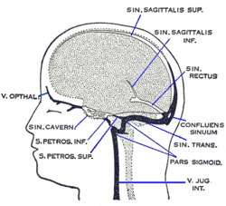
The cavernous sinus within the human head is one of the dural venous sinuses creating a cavity called the lateral sellar compartment bordered by the temporal bone of the skull and the sphenoid bone, lateral to the sella turcica.

The dural venous sinuses are venous sinuses (channels) found between the endosteal and meningeal layers of dura mater in the brain. They receive blood from the cerebral veins, and cerebrospinal fluid (CSF) from the subarachnoid space via arachnoid granulations. They mainly empty into the internal jugular vein. Cranial venous sinuses communicate with veins outside the skull through emissary veins. These communications help to keep the pressure of blood in the sinuses constant. The major dural venous sinuses included the superior sagittal sinus, inferior sagittal sinus, transverse sinus, straight sinus, sigmoid sinus and cavernous sinus. These sinuses play a crucial role in cerebral venous drainage. A dural venous sinus, in human anatomy, is any of the channels of a branching complex sinus network that lies between layers of the dura mater, the outermost covering of the brain, and functions to collect oxygen-depleted blood. Unlike veins, these sinuses possess no muscular coat.
The confluence of sinuses, torcular Herophili, or torcula is the connecting point of the superior sagittal sinus, straight sinus, and occipital sinus. It is below the internal occipital protuberance of the skull. It drains venous blood from the brain into the transverse sinuses. It may be affected by arteriovenous fistulas, a thrombus, major trauma, or surgical damage, and may be imaged with many radiology techniques.

The occipital sinus is the smallest of the dural venous sinuses. It is usually unpaired, and is sometimes altogether absent. It is situated in the attached margin of the falx cerebelli. It commences near the foramen magnum, and ends by draining into the confluence of sinuses.
The superior sagittal sinus, within the human head, is an unpaired area along the attached margin of the falx cerebri. It allows blood to drain from the lateral aspects of anterior cerebral hemispheres to the confluence of sinuses. Cerebrospinal fluid drains through arachnoid granulations into the superior sagittal sinus and is returned to venous circulation.
The inferior sagittal sinus, within the human head, is an area beneath the brain which allows blood to drain outwards posteriorly from the center of the head. It drains to the straight sinus, which connects to the transverse sinuses. See diagram : labeled in the brain as "SIN. SAGITTALIS INF.".
The superior petrosal sinus is one of the dural venous sinuses located beneath the brain. It receives blood from the cavernous sinus and passes backward and laterally to drain into the transverse sinus. The sinus receives superior petrosal veins, some cerebellar veins, some inferior cerebral veins, and veins from the tympanic cavity. They may be affected by arteriovenous malformation or arteriovenous fistula, usually treated with surgery.
The falx cerebelli is a small sickle-shaped fold of dura mater projecting forwards into the posterior cerebellar notch as well as projecting into the vallecula of the cerebellum between the two cerebellar hemispheres.
The sigmoid sinuses, also known as the pars sigmoid, are paired dural venous sinuses within the skull that receive blood from posterior transverse sinuses.
The transverse sinuses, within the human head, are two areas beneath the brain which allow blood to drain from the back of the head. They run laterally in a groove along the interior surface of the occipital bone. They drain from the confluence of sinuses to the sigmoid sinuses, which ultimately connect to the internal jugular vein. See diagram : labeled under the brain as "SIN. TRANS.".

The squamous part of occipital bone is situated above and behind the foramen magnum, and is curved from above downward and from side to side.

The cerebellar veins are veins which drain the cerebellum. They consist of the superior cerebellar veins and the inferior cerebellar veins. The superior cerebellar veins drain to the straight sinus and the internal cerebral veins. The inferior cerebellar veins drain to the transverse sinus, the superior petrosal sinus, and the occipital sinus.

A falcine sinus is a venous channel that lies within the falx cerebri connecting the vein of Galen and the posterior part of superior sagittal sinus. It is normally present during fetal development and involutes after birth. The presence of a falcine sinus has been associated with a vein of Galen malformation and other vascular anomalies. The persistence of a falcine sinus after the neonatal period was previously thought to be rare, but has recently been described to be present in up to 5% of all people, appearings in approximately 2.1% of CT examinations of adult patients. Some authors have studied the plexus rather than the sinus, a rare form of the venous pathway between the layers of the cerebral falx, which connects the superior sagittal sinus with the inferior sagittal sinus and the straight sinus.