
At the base of the skull, the foramen ovale is one of the larger of the several holes that transmit nerves through the skull. The foramen ovale is situated in the posterior part of the sphenoid bone, posterolateral to the foramen rotundum.

The scalp is the anatomical area bordered by the human face at the front, and by the neck at the sides and back.

The pulmonary veins are the veins that transfer oxygenated blood from the lungs to the heart. The largest pulmonary veins are the four main pulmonary veins, two from each lung that drain into the left atrium of the heart. The pulmonary veins are part of the pulmonary circulation.
The emissary veins connect the extracranial venous system with the intracranial venous sinuses. They connect the veins outside the cranium to the venous sinuses inside the cranium. They drain from the scalp, through the skull, into the larger meningeal veins and dural venous sinuses.

The cavernous sinus within the human head, is one of the dural venous sinuses creating a cavity called the lateral sellar compartment bordered by the temporal bone of the skull and the sphenoid bone, lateral to the sella turcica.
The superior sagittal sinus, within the human head, is an unpaired area along the attached margin of the falx cerebri. It allows blood to drain from the lateral aspects of anterior cerebral hemispheres to the confluence of sinuses. Cerebrospinal fluid drains through arachnoid granulations into the superior sagittal sinus and is returned to venous circulation.
The sigmoid sinuses, also known as the pars sigmoid, are venous sinuses within the skull that receive blood from posterior dural venous sinus veins.
The transverse sinuses, within the human head, are two areas beneath the brain which allow blood to drain from the back of the head. They run laterally in a groove along the interior surface of the occipital bone. They drain from the confluence of sinuses to the sigmoid sinuses, which ultimately connect to the internal jugular vein. See diagram : labeled under the brain as "SIN. TRANS.".
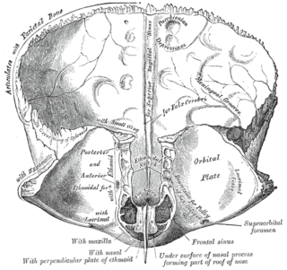
The frontal crest of the frontal bone ends below in a small notch which is converted into a foramen, the foramen cecum, by articulation with the ethmoid.

The occipital vein begins as a plexus at the posterior aspect of the scalp from the external occipital protuberance and superior nuchal line to the back part of the vertex of the skull.
In the base of the skull, in the great wings of the sphenoid bone, medial to the foramen ovale, a small aperture, the sphenoidal emissary foramen, may occasionally be seen opposite the root of the pterygoid process. When present, it opens below near the scaphoid fossa. Vesalius was the first to describe and illustrate this foramen, and it thus sometimes bears the name of foramen Vesalii. Other names include foramen venosum and canaliculus sphenoidalis.

The petrosquamous suture is a cranial suture between the petrous portion and the squama of the temporal bone. It forms the Koerner's septum. The petrous portion forms the medial component of the osseous margin, while the squama forms the lateral component. The anterolateral portion (squama) arises from the mesenchyme at 8 weeks of embryogenesis while the petromastoid portion develops later from a cartilaginous center at 6 months of fetal development.

The mastoid foramen is a hole in the posterior border of the temporal bone. It transmits a Mastoid emissary vein to the sigmoid sinus and a small branch of the occipital artery, the posterior meningeal artery to the dura mater.

The nerve of the pterygoid canal is formed by the junction of the greater petrosal nerve and the deep petrosal nerve within the pterygoid canal containing the cartilaginous substance, which fills the foramen lacerum.
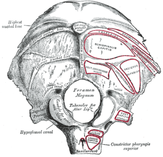
Behind either condyle of the lateral parts of occipital bone is a depression, the condyloid fossa, which receives the posterior margin of the superior facet of the atlas when the head is bent backward; the floor of this fossa is sometimes perforated by the condyloid canal, through which an emissary vein passes from the transverse sinus.

The parietal foramen is an opening for the parietal emissary vein, which drains into the superior sagittal sinus. Occasionally, a small branch of the occipital artery can also pass through it. It is located at the back part of the parietal bone, close to the upper or sagittal border. It is not always present, and its size varies considerably.

The internal vertebral venous plexuses lie within the vertebral canal in the epidural space, and receive tributaries from the bones and from the spinal cord.

In human anatomy, the dorsal veins of the penis comprise the superficial dorsal vein of the penis and the deep dorsal vein of the penis.
The public domain consists of all the creative works to which no exclusive intellectual property rights apply. Those rights may have expired, been forfeited, expressly waived, or may be inapplicable.

Gray's Anatomy is an English language textbook of human anatomy originally written by Henry Gray and illustrated by Henry Vandyke Carter. Earlier editions were called Anatomy: Descriptive and Surgical, Anatomy of the Human Body and Gray's Anatomy: Descriptive and Applied, but the book's name is commonly shortened to, and later editions are titled, Gray's Anatomy. The book is widely regarded as an extremely influential work on the subject, and has continued to be revised and republished from its initial publication in 1858 to the present day. The latest edition of the book, the 41st, was published in September 2015.