
The third ventricle is one of the four connected ventricles of the ventricular system within the mammalian brain. It is a slit-like cavity formed in the diencephalon between the two thalami, in the midline between the right and left lateral ventricles, and is filled with cerebrospinal fluid (CSF).

The internal carotid artery is located in the inner side of the neck in contrast to the external carotid artery. In human anatomy, they arise from the common carotid arteries where these bifurcate into the internal and external carotid arteries at cervical vertebral level 3 or 4; the internal carotid artery supplies the brain including eyes, while the external carotid nourishes other portions of the head, such as the face, scalp, skull, and meninges.

The sella turcica is a saddle-shaped depression in the body of the sphenoid bone of the human skull and of the skulls of other hominids including chimpanzees, orangutans and gorillas. It serves as a cephalometric landmark. The pituitary gland or hypophysis is located within the most inferior aspect of the sella turcica, the hypophyseal fossa.

Dura mater is a thick membrane made of dense irregular connective tissue that surrounds the brain and spinal cord. It is the outermost of the three layers of membrane called the meninges that protect the central nervous system. The other two meningeal layers are the arachnoid mater and the pia mater. The dura surrounds the brain and the spinal cord. It envelops the arachnoid mater, which is responsible for keeping in the cerebrospinal fluid. It is derived primarily from the neural crest cell population, with postnatal contributions of the paraxial mesoderm.

The posterior cranial fossa is part of the cranial cavity, located between the foramen magnum and tentorium cerebelli. It contains the brainstem and cerebellum.

The ophthalmic artery (OA) is the first branch of the internal carotid artery distal to the cavernous sinus. Branches of the OA supply all the structures in the orbit as well as some structures in the nose, face and meninges. Occlusion of the OA or its branches can produce sight-threatening conditions.
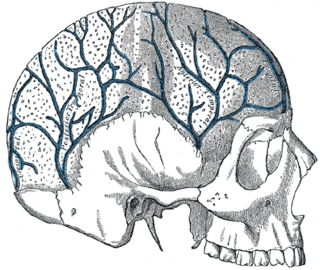
The diploic veins are large, thin-walled valveless veins that channel in the diploë between the inner and outer layers of the cortical bone in the skull. They are lined by a single layer of endothelium supported by elastic tissue. They develop fully by the age of two years. The diploic veins drain this area into the dural venous sinuses. The four major trunks of the diploic veins found on each side of the head are frontal, anterior temporal, posterior temporal, and occipital diploic veins.

The cavernous sinus within the human head is one of the dural venous sinuses creating a cavity called the lateral sellar compartment bordered by the temporal bone of the skull and the sphenoid bone, lateral to the sella turcica.
The superior sagittal sinus, within the human head, is an unpaired area along the attached margin of the falx cerebri. It allows blood to drain from the lateral aspects of anterior cerebral hemispheres to the confluence of sinuses. Cerebrospinal fluid drains through arachnoid granulations into the superior sagittal sinus and is returned to venous circulation.
The inferior petrosal sinuses are two small sinuses situated on the inferior border of the petrous part of the temporal bone, one on each side. Each inferior petrosal sinus drains the cavernous sinus into the internal jugular vein.

The superior ophthalmic vein begins at the inner angle of the orbit in a vein named the nasofrontal which communicates anteriorly with the angular vein; it does not pursue the same course as the ophthalmic artery and receives tributaries corresponding to the branches of that vessel.

The sphenoid sinus is one of the four paired paranasal sinuses that is contained within the body of the sphenoid bone. The sphenoid sinuses vary in size and shape, and owing to the lateral displacement of the intervening septum, which may insert on the carotid canal, they are rarely symmetrical. They cannot be palpated during an extraoral examination.

The facial vein is a relatively large vein in the human face. It commences at the side of the root of the nose and is a direct continuation of the angular vein where it also receives a small nasal branch. It lies behind the facial artery and follows a less tortuous course. It receives blood from the external palatine vein before it either joins the anterior branch of the retromandibular vein to form the common facial vein, or drains directly into the internal jugular vein.

The pterygoid plexus is a venous plexus of considerable size, and is situated between the temporalis muscle and lateral pterygoid muscle, and partly between the two pterygoid muscles.

The middle cranial fossa, deeper than the anterior cranial fossa, is narrow medially and widens laterally to the sides of the skull. It is separated from the posterior fossa by the clivus and the petrous crest.

In the sphenoid bone, the anterior boundary of the sella turcica is completed by two small eminences, one on either side, called the anterior clinoid processes, while the posterior boundary is formed by a square-shaped plate of bone, the dorsum sellæ, ending at its superior angles in two tubercles, the posterior clinoid processes, the size and form of which vary considerably in different individuals. The posterior clinoid processes deepen the sella turcica, and give attachment to the tentorium cerebelli.

The internal vertebral venous plexuses lie within the vertebral canal in the epidural space, and receive tributaries from the bones and from the spinal cord.

The spinal veins are situated in the pia mater and form a minute, tortuous, venous plexus.
The central retinal vein is a short vein that runs through the optic nerve, leaves the optic nerve 10 mm from the eyeball and drains blood from the capillaries of the retina into either superior ophthalmic vein or into the cavernous sinus directly. The anatomy of the veins of the orbit of the eye varies between individuals, and in some the central retinal vein drains into the superior ophthalmic vein, and in some it drains directly into the cavernous sinus.
Anterior spinal veins are veins that receive blood from the anterior spinal cord.


