Related Research Articles

The scalp is the area of the head where head hair grows. It is made up of skin, layers of connective and fibrous tissues, and the membrane of the skull. Anatomically, the scalp is part of the epicranium, a collection of structures covering the cranium. The scalp is bordered by the face at the front, and by the neck at the sides and back. The scientific study of hair and scalp is called trichology.

The facial artery is a branch of the external carotid artery that supplies structures of the superficial face.

In human anatomy, the superficial temporal artery is a major artery of the head. It arises from the external carotid artery when it splits into the superficial temporal artery and maxillary artery.

The cavernous sinus within the human head is one of the dural venous sinuses creating a cavity called the lateral sellar compartment bordered by the temporal bone of the skull and the sphenoid bone, lateral to the sella turcica.

The superior ophthalmic vein is a vein of the orbit that drains venous blood from structures of the upper orbit. It is formed by the union of the angular vein, and supraorbital vein. It passes backwards within the orbit alongside the ophthalmic artery, then exits the orbit through the superior orbital fissure to drain into the cavernous sinus.

The facial vein is a relatively large vein in the human face. It commences at the side of the root of the nose and is a direct continuation of the angular vein where it also receives a small nasal branch. It lies behind the facial artery and follows a less tortuous course. It receives blood from the external palatine vein before it either joins the anterior branch of the retromandibular vein to form the common facial vein, or drains directly into the internal jugular vein.
The facial vein usually unites with the anterior branch of the retromandibular vein to form the common facial vein, which crosses the external carotid artery and enters the internal jugular vein at a variable point below the hyoid bone.
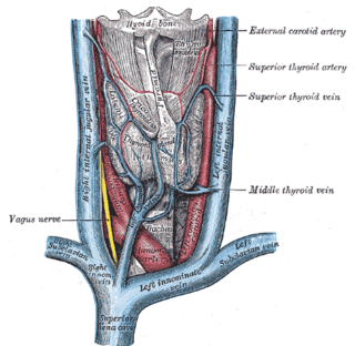
The superior thyroid vein is the vena comitans of the superior thyroid artery. It is formed by the union of deep and superficial tributaries that correspond to the arterial branches of the superior thyroid artery. Its tributaries are the superior laryngeal vein, and the cricothyroid veins. The vein empties into either the internal jugular vein, or the facial vein.
The maxillary vein or internal maxillary vein is a vein of the head. It is a short trunk which accompanies the maxillary artery. It is formed by a confluence of the veins of the pterygoid plexus. It and passes posterior-ward between the sphenomandibular ligament and the neck of the mandible to enter the parotid gland where unites with the superficial temporal vein to form the retromandibular vein.

The pterygoid plexus is a fine venous plexus upon and within the lateral pterygoid muscle. It drains by a short maxillary vein.

The transverse facial artery is an artery that branches from the superficial temporal artery and runs across the face.
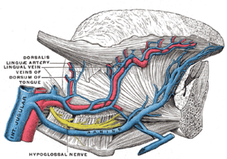
The lingual veins are multiple veins of the tongue with two distinct courses: one group drains into the lingual artery; another group drains either into the lingual artery, (common) facial vein, or internal jugular vein.
The superficial temporal vein is a vein of the side of the head which collects venous blood from the region of the temple. It arises from an anastomosing venous plexus on the side and vertex of the head. The superficial temporal vein terminates within the substance of the parotid gland by uniting with the maxillary vein to form the retromandibular vein.
The angular vein is a vein of the face. It is the upper part of the facial vein, above its junction with the superior labial vein. It is formed by the junction of the supratrochlear vein and supraorbital vein, and joins with the superior labial vein. It drains the medial canthus, and parts of the nose and the upper lip. It can be a route of spread of infection from the danger triangle of the face to the cavernous sinus.
The retromandibular vein is a major vein of the face. It is formed within the parotid gland by the confluence of the maxillary vein, and superficial temporal vein. It descends in the gland and splits into two branches upon emerging from the gland. Its anterior branch then joins the (anterior) facial vein forming the common facial vein, while its posterior branch joins the posterior auricular vein forming the external jugular vein.
The pharyngeal veins commence in the pharyngeal plexus superficial to the pharynx. The pharyngeal veins receive as tributaries meningeal vein, and the vein of the pterygoid canal. The pharyngeal veins typically empty into the internal jugular vein.

The marginal mandibular branch of the facial nerve arises from the facial nerve in the parotid gland at the parotid plexus. It passes anterior-ward deep to the platysma and depressor anguli oris muscles. It provides motor innervation to muscles of the lower lip and chin: the depressor labii inferioris muscle, depressor anguli oris muscle, and mentalis muscle. It communicates with the mental branch of the inferior alveolar nerve.

The submandibular triangle corresponds to the region of the neck immediately beneath the body of the mandible.

The carotid triangle is a portion of the anterior triangle of the neck.
The deep lingual vein is one of the lingual veins. It commences near the apex of the tongue. It passes posterior-ward close to the inferior surface of the tongue. It terminates near the anterior border of the hyoglossus muscle by uniting with the sublingual vein to form the vena comitans of the hypoglossal nerve ; this vein then passes posterior-ward alongside the nerve to empty into either a lingual vein, the (common) facial vein, or the internal jugular vein.
References
![]() This article incorporates text in the public domain from page 645 of the 20th edition of Gray's Anatomy (1918)
This article incorporates text in the public domain from page 645 of the 20th edition of Gray's Anatomy (1918)