
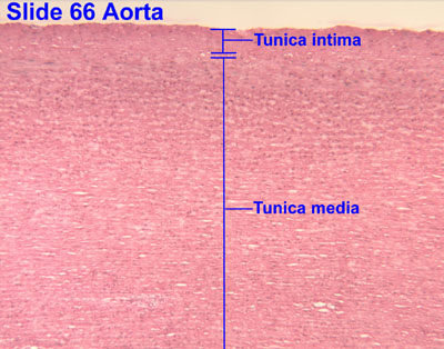
The aorta is the main and largest artery in the human body, originating from the left ventricle of the heart, branching upwards immediately after, and extending down to the abdomen, where it splits at the aortic bifurcation into two smaller arteries. The aorta distributes oxygenated blood to all parts of the body through the systemic circulation.

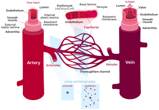
An artery is a blood vessel in humans and most other animals that takes oxygenated blood away from the heart in the systemic circulation to one or more parts of the body. Exceptions that carry deoxygenated blood are the pulmonary arteries in the pulmonary circulation that carry blood to the lungs for oxygenation, and the umbilical arteries in the fetal circulation that carry deoxygenated blood to the placenta. It consists of a multi-layered artery wall wrapped into a tube-shaped channel.
Blood vessels are the tubular structures of a circulatory system that transport blood throughout a vertebrate's body. Blood vessels transport blood cells, nutrients, and oxygen to most of the tissues of a body. They also take waste and carbon dioxide away from the tissues. Some tissues such as cartilage, epithelium, and the lens and cornea of the eye are not supplied with blood vessels and are termed avascular.

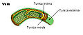
Veins are blood vessels in the circulatory system of humans and most other animals that carry blood towards the heart. Most veins carry deoxygenated blood from the tissues back to the heart; exceptions are those of the pulmonary and fetal circulations which carry oxygenated blood to the heart. In the systemic circulation, arteries carry oxygenated blood away from the heart, and veins return deoxygenated blood to the heart, in the deep veins.

The ileum is the final section of the small intestine in most higher vertebrates, including mammals, reptiles, and birds. In fish, the divisions of the small intestine are not as clear and the terms posterior intestine or distal intestine may be used instead of ileum. Its main function is to absorb vitamin B12, bile salts, and whatever products of digestion that were not absorbed by the jejunum.

The superior vena cava (SVC) is the superior of the two venae cavae, the great venous trunks that return deoxygenated blood from the systemic circulation to the right atrium of the heart. It is a large-diameter (24 mm) short length vein that receives venous return from the upper half of the body, above the diaphragm. Venous return from the lower half, below the diaphragm, flows through the inferior vena cava. The SVC is located in the anterior right superior mediastinum. It is the typical site of central venous access via a central venous catheter or a peripherally inserted central catheter. Mentions of "the cava" without further specification usually refer to the SVC.

A venule is a very small vein in the microcirculation that allows blood to return from the capillary beds to drain into the venous system via increasingly larger veins. Post-capillary venules are the smallest of the veins with a diameter of between 10 and 30 micrometres (μm). When the post-capillary venules increase in diameter to 50μm they can incorporate smooth muscle and are known as muscular venules. Veins contain approximately 70% of total blood volume, while about 25% is contained in the venules. Many venules unite to form a vein.

Vasa vasorum are networks of small blood vessels that supply the walls of large blood vessels, such as elastic arteries and large veins.
The basal lamina is a layer of extracellular matrix secreted by the epithelial cells, on which the epithelium sits. It is often incorrectly referred to as the basement membrane, though it does constitute a portion of the basement membrane. The basal lamina is visible only with the electron microscope, where it appears as an electron-dense layer that is 20–100 nm thick.

The basement membrane, also known as base membrane, is a thin, pliable sheet-like type of extracellular matrix that provides cell and tissue support and acts as a platform for complex signalling. The basement membrane sits between epithelial tissues including mesothelium and endothelium, and the underlying connective tissue.

The adventitia is the outer layer of fibrous connective tissue surrounding an organ.

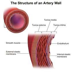
The tunica intima, or intima for short, is the innermost tunica (layer) of an artery or vein. It is made up of one layer of endothelial cells, and is supported by an internal elastic lamina. The endothelial cells are in direct contact with the blood flow.
In cardiology, the cardiac skeleton, also known as the fibrous skeleton of the heart, is a high-density homogeneous structure of connective tissue that forms and anchors the valves of the heart, and influences the forces exerted by and through them. The cardiac skeleton separates and partitions the atria from the ventricles. The heart's cardiac skeleton comprises four dense connective tissue rings that encircle the mitral and tricuspid atrioventricular (AV) canals and extend to the origins of the pulmonary trunk and aorta. This provides crucial support and structure to the heart while also serving to electrically isolate the atria from the ventricles.
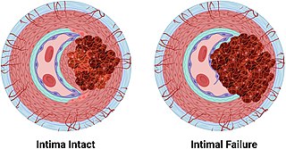
An arterial dissection is a tear within the wall of an artery, which allows blood to separate the wall layers. There are several types. Tears almost always occur in arterial walls, but a vein wall tear has been documented.

The muscular layer is a region of muscle in many organs in the vertebrate body, adjacent to the submucosa. It is responsible for gut movement such as peristalsis. The Latin, tunica muscularis, may also be used.
The tunica albuginea is the fibrous envelope that extends the length of the corpus cavernosum penis and corpus spongiosum penis. It is a bi-layered structure that includes an outer longitudinal layer and an inner circular layer.
The tunica externa, also known as the tunica adventitia, is the outermost tunica (layer) of a blood vessel, surrounding the tunica media. It is mainly composed of collagen and, in arteries, is supported by external elastic lamina. The collagen serves to anchor the blood vessel to nearby organs, giving it stability.
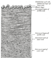
An elastic artery is an artery with many collagen and elastin filaments in the tunica media, which gives it the ability to stretch in response to each pulse. This elasticity also gives rise to the Windkessel effect, which helps to maintain a relatively constant pressure in the arteries despite the pulsating nature of the blood flow. Elastic arteries include the largest arteries in the body, those closest to the heart. They give rise to medium-sized vessels known as distributing arteries.
A muscular artery is a medium-sized artery that draws blood from an elastic artery and branches into "resistance vessels" including small arteries and arterioles. Their walls contain larger number of smooth muscles, allowing them to contract and expand depending on peripheral blood demand.

The internal elastic lamina or internal elastic lamella is a layer of elastic tissue that forms the outermost part of the tunica intima of blood vessels. It separates tunica intima from tunica media.


