Related Research Articles

The peripheral nervous system (PNS) is one of two components that make up the nervous system of bilateral animals, with the other part being the central nervous system (CNS). The PNS consists of nerves and ganglia, which lie outside the brain and the spinal cord. The main function of the PNS is to connect the CNS to the limbs and organs, essentially serving as a relay between the brain and spinal cord and the rest of the body. Unlike the CNS, the PNS is not protected by the vertebral column and skull, or by the blood–brain barrier, which leaves it exposed to toxins.

Cranial nerves are the nerves that emerge directly from the brain, of which there are conventionally considered twelve pairs. Cranial nerves relay information between the brain and parts of the body, primarily to and from regions of the head and neck, including the special senses of vision, taste, smell, and hearing.

The pudendal nerve is the main nerve of the perineum. It is a mixed nerve and also conveys sympathetic autonomic fibers. It carries sensation from the external genitalia of both sexes and the skin around the anus and perineum, as well as the motor supply to various pelvic muscles, including the male or female external urethral sphincter and the external anal sphincter.

The sympathetic nervous system is one of the three divisions of the autonomic nervous system, the others being the parasympathetic nervous system and the enteric nervous system. The enteric nervous system is sometimes considered part of the autonomic nervous system, and sometimes considered an independent system.

A spinal nerve is a mixed nerve, which carries motor, sensory, and autonomic signals between the spinal cord and the body. In the human body there are 31 pairs of spinal nerves, one on each side of the vertebral column. These are grouped into the corresponding cervical, thoracic, lumbar, sacral and coccygeal regions of the spine. There are eight pairs of cervical nerves, twelve pairs of thoracic nerves, five pairs of lumbar nerves, five pairs of sacral nerves, and one pair of coccygeal nerves. The spinal nerves are part of the peripheral nervous system.
Paresthesia is an abnormal sensation of the skin with no apparent physical cause. Paresthesia may be transient or chronic, and may have many possible underlying causes. Paresthesias are usually painless and can occur anywhere on the body, but most commonly occur in the arms and legs.
Diabetic neuropathy includes various types of nerve damage associated with diabetes mellitus. The most common form, diabetic peripheral neuropathy, affects 30% of all diabetic patients. Symptoms depend on the site of nerve damage and can include motor changes such as weakness; sensory symptoms such as numbness, tingling, or pain; or autonomic changes such as urinary symptoms. These changes are thought to result from a microvascular injury involving small blood vessels that supply nerves. Relatively common conditions which may be associated with diabetic neuropathy include distal symmetric polyneuropathy; third, fourth, or sixth cranial nerve palsy; mononeuropathy; mononeuropathy multiplex; diabetic amyotrophy; and autonomic neuropathy.
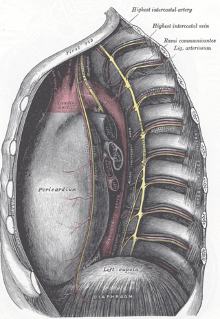
The ligamentum arteriosum, also known as Botallo's ligament, Harvey's ligament, and Botallo's duct, is a small ligament attaching the aorta to the pulmonary artery. It serves no function in adults but is the remnant of the ductus arteriosus formed within three weeks after birth.
Leukocytosis is a condition in which the white cell (leukocyte) count is above the normal range in the blood. It is frequently a sign of an inflammatory response, most commonly the result of infection, but may also occur following certain parasitic infections or bone tumors as well as leukemia. It may also occur after strenuous exercise, convulsions such as epilepsy, emotional stress, pregnancy and labor, anesthesia, as a side effect of medication, and epinephrine administration. There are five principal types of leukocytosis:
- Neutrophilia
- Lymphocytosis
- Monocytosis
- Eosinophilia
- Basophilia

Vasa vasorum are small blood vessels that comprise a vascular network supplying the walls of large blood vessels, such as elastic arteries and large veins.

Polyarteritis nodosa (PAN) is a systemic necrotizing inflammation of blood vessels (vasculitis) affecting medium-sized muscular arteries, typically involving the arteries of the kidneys and other internal organs but generally sparing the lungs' circulation. Small aneurysms are strung like the beads of a rosary, therefore making this "rosary sign" an important diagnostic feature of the vasculitis. PAN is sometimes associated with infection by the hepatitis B or hepatitis C virus. The condition may be present in infants.

Neuritis, from the Greek νεῦρον), is inflammation of a nerve or the general inflammation of the peripheral nervous system. Inflammation, and frequently concomitant demyelination, cause impaired transmission of neural signals and leads to aberrant nerve function. Neuritis is often conflated with neuropathy, a broad term describing any disease process which affects the peripheral nervous system. However, neuropathies may be due to either inflammatory or non-inflammatory causes, and the term encompasses any form of damage, degeneration, or dysfunction, while neuritis refers specifically to the inflammatory process.

The inferior gluteal nerve is the main motor neuron that innervates the gluteus maximus muscle. It is responsible for the movement of the gluteus maximus in activities requiring the hip to extend the thigh, such as climbing stairs. Injury to this nerve is rare but often occurs as a complication of posterior approach to the hip during hip replacement. When damaged, one would develop gluteus maximus lurch, which is a gait abnormality which causes the individual to 'lurch' backwards to compensate lack in hip extension.
The superior gluteal nerve is a mixed nerve of the sacral plexus that originates in the pelvis. It provides motor innervation to the gluteus medius, gluteus minimus, tensor fasciae latae, and piriformis muscles; it also has a cutaneous branch.
Lippincott Williams & Wilkins (LWW) is an American imprint of the American Dutch publishing conglomerate Wolters Kluwer. It was established by the acquisition of Williams & Wilkins and its merger with J.B. Lippincott Company in 1998. Under the LWW brand, Wolters Kluwer, through its Health Division, publishes scientific, technical, and medical content such as textbooks, reference works, and over 275 scientific journals. Publications are aimed at physicians, nurses, clinicians, and students.
Toxic and nutritional optic neuropathy is a group of medical disorders defined by visual impairment due to optic nerve damage secondary to a toxic substance and/or nutritional deficiency. The causes of these disorders are various, but they are linked by shared signs and symptoms, which this article will describe. In several of these disorders, both toxic and nutritional factors play a role, acting synergistically.
Biceps reflex is a deep tendon reflex (DTR) test that examines the function of the C5 reflex arc and the C6 reflex arc. The test is performed by using a tendon hammer to quickly depress the biceps brachii tendon as it passes through the cubital fossa. Specifically, the test activates the stretch receptors inside the biceps brachii muscle which communicates mainly with the C5 spinal nerve and partially with the C6 spinal nerve to induce a reflex contraction of the biceps muscle and jerk of the forearm.

The arcuate vessels of the uterus are a component of the blood supply of the uterus. They are arteries and veins that branch from the uterine arteries and veins, respectively, with additional anastomoses from the ovarian arteries and veins, and penetrate and assume a circumferential course in the myometrium.

Intercostal nerve block is a nerve block which temporarily or permanently interrupts the flow of signals along an intercostal nerve, usually performed to relieve pain.

Shin Joong Oh is a Korean physician who is Distinguished Professor of Neurology Emeritus at The University of Alabama at Birmingham in the United States. Oh is a clinician, researcher, and educator known for his contributions to the fields of neurology and electrodiagnostic medicine, particularly electromyography. He retired in 2014.
References
- ↑ Rohkamm, Reinhard (2004). Color atlas of neurology . Ill. by Manfred Güther. Transl. rev. by Ethan Taub. [Orig. transl.: Suzyon O'Neal Wandrey]. Stuttgart [u.a.]: Thieme. pp. 90. ISBN 9781588901910.
- 1 2 Moore, Keith L.; Agur, Anne M.R.; Dalley, Arthur F. (2010). Clinically oriented anatomy (6th ed., [International ed.]. ed.). Philadelphia [etc.]: Lippincott Williams & Wilkins, Wolters Kluwer. p. 50. ISBN 9781605476520.
- 1 2 3 Imboden, John B.; Stone, John H.; Hellman, David B., eds. (2007). Current rheumatology diagnosis & treatment (2nd ed.). New York; Toronto: Lange Medical Books/McGraw-Hill, Medical Pub. Division. ISBN 9780071460408.
- ↑ Said, G (November 1997). "Necrotizing peripheral nerve vasculitis". Neurologic Clinics. 15 (4): 835–48. doi:10.1016/s0733-8619(05)70350-9. PMID 9367967.
- ↑ Chelly, Jacques E., ed. (2009). Peripheral nerve blocks : a color atlas (3rd ed.). Philadelphia, PA: Wolters Kluwer Health/Lippincott Williams & Wilkins. p. 214. ISBN 9780781768764.