In neuroscience and psychology, the term language center refers collectively to the areas of the brain which serve a particular function for speech processing and production. Language is a core system that gives humans the capacity to solve difficult problems and provides them with a unique type of social interaction. Language allows individuals to attribute symbols to specific concepts, and utilize them through sentences and phrases that follow proper grammatical rules. Finally, speech is the mechanism by which language is orally expressed.

Wernicke's aphasia, also known as receptive aphasia, sensory aphasia, fluent aphasia, or posterior aphasia, is a type of aphasia in which individuals have difficulty understanding written and spoken language. Patients with Wernicke's aphasia demonstrate fluent speech, which is characterized by typical speech rate, intact syntactic abilities and effortless speech output. Writing often reflects speech in that it tends to lack content or meaning. In most cases, motor deficits do not occur in individuals with Wernicke's aphasia. Therefore, they may produce a large amount of speech without much meaning. Individuals with Wernicke's aphasia often suffer of anosognosia – they are unaware of their errors in speech and do not realize their speech may lack meaning. They typically remain unaware of even their most profound language deficits.

Broca's area, or the Broca area, is a region in the frontal lobe of the dominant hemisphere, usually the left, of the brain with functions linked to speech production.
Aphasiology is the study of language impairment usually resulting from brain damage, due to neurovascular accident—hemorrhage, stroke—or associated with a variety of neurodegenerative diseases, including different types of dementia. These specific language deficits, termed aphasias, may be defined as impairments of language production or comprehension that cannot be attributed to trivial causes such as deafness or oral paralysis. A number of aphasias have been described, but two are best known: expressive aphasia and receptive aphasia.

A Brodmann area is a region of the cerebral cortex, in the human or other primate brain, defined by its cytoarchitecture, or histological structure and organization of cells. The concept was first introduced by the German anatomist Korbinian Brodmann in the early 20th century. Brodmann mapped the human brain based on the varied cellular structure across the cortex and identified 52 distinct regions, which he numbered 1 to 52. These regions, or Brodmann areas, correspond with diverse functions including sensation, motor control, and cognition.

The temporal lobe is one of the four major lobes of the cerebral cortex in the brain of mammals. The temporal lobe is located beneath the lateral fissure on both cerebral hemispheres of the mammalian brain.

Brodmann area 47, or BA47, is part of the frontal cortex in the human brain. It curves from the lateral surface of the frontal lobe into the ventral (orbital) frontal cortex. It is below areas BA10 and BA45, and beside BA11. This cytoarchitectonic region most closely corresponds to the gyral region the orbital part of inferior frontal gyrus, although these regions are not equivalent. Pars orbitalis is not based on cytoarchitectonic distinctions, and rather is defined according to gross anatomical landmarks. Despite a clear distinction, these two terms are often used liberally in peer-reviewed research journals.
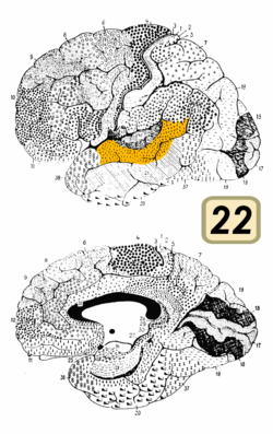
Wernicke's area, also called Wernicke's speech area, is one of the two parts of the cerebral cortex that are linked to speech, the other being Broca's area. It is involved in the comprehension of written and spoken language, in contrast to Broca's area, which is primarily involved in the production of language. It is traditionally thought to reside in Brodmann area 22, which is located in the superior temporal gyrus in the dominant cerebral hemisphere, which is the left hemisphere in about 95% of right-handed individuals and 70% of left-handed individuals.
Conduction aphasia, also called associative aphasia, is an uncommon form of difficulty in speaking (aphasia). It is caused by damage to the parietal lobe of the brain. An acquired language disorder, it is characterised by intact auditory comprehension, coherent speech production, but poor speech repetition. Affected people are fully capable of understanding what they are hearing, but fail to encode phonological information for production. This deficit is load-sensitive as the person shows significant difficulty repeating phrases, particularly as the phrases increase in length and complexity and as they stumble over words they are attempting to pronounce. People have frequent errors during spontaneous speech, such as substituting or transposing sounds. They are also aware of their errors and will show significant difficulty correcting them.

The inferior frontal gyrus (IFG),, is the lowest positioned gyrus of the frontal gyri, of the frontal lobe, and is part of the prefrontal cortex.

The transverse temporal gyri, also called Heschl's gyri or Heschl's convolutions, are gyri found in the area of primary auditory cortex buried within the lateral sulcus of the human brain, occupying Brodmann areas 41 and 42. Transverse temporal gyri are superior to and separated from the planum temporale by Heschl's sulcus. Transverse temporal gyri are found in varying numbers in both the right and left hemispheres of the brain and one study found that this number is not related to the hemisphere or dominance of hemisphere studied in subjects. Transverse temporal gyri can be viewed in the sagittal plane as either an omega shape or a heart shape.

The superior temporal gyrus (STG) is one of three gyri in the temporal lobe of the human brain, which is located laterally to the head, situated somewhat above the external ear.

In neuroanatomy, the primary somatosensory cortex is located in the postcentral gyrus of the brain's parietal lobe, and is part of the somatosensory system. It was initially defined from surface stimulation studies of Wilder Penfield, and parallel surface potential studies of Bard, Woolsey, and Marshall. Although initially defined to be roughly the same as Brodmann areas 3, 1 and 2, more recent work by Kaas has suggested that for homogeny with other sensory fields only area 3 should be referred to as "primary somatosensory cortex", as it receives the bulk of the thalamocortical projections from the sensory input fields.

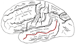
In neuroanatomy, the arcuate fasciculus is a bundle of axons that generally connects the Broca's area and the Wernicke's area in the brain. It is an association fiber tract connecting caudal temporal cortex and inferior frontal lobe.
Transcortical sensory aphasia (TSA) is a kind of aphasia that involves damage to specific areas of the temporal lobe of the brain, resulting in symptoms such as poor auditory comprehension, relatively intact repetition, and fluent speech with semantic paraphasias present. TSA is a fluent aphasia similar to Wernicke's aphasia, with the exception of a strong ability to repeat words and phrases. The person may repeat questions rather than answer them ("echolalia").

In psycholinguistics, language processing refers to the way humans use words to communicate ideas and feelings, and how such communications are processed and understood. Language processing is considered to be a uniquely human ability that is not produced with the same grammatical understanding or systematicity in even human's closest primate relatives.
Auditory agnosia is a form of agnosia that manifests itself primarily in the inability to recognize or differentiate between sounds. It is not a defect of the ear or "hearing", but rather a neurological inability of the brain to process sound meaning. While auditory agnosia impairs the understanding of sounds, other abilities such as reading, writing, and speaking are not hindered. It is caused by bilateral damage to the anterior superior temporal gyrus, which is part of the auditory pathway responsible for sound recognition, the auditory "what" pathway.
In the human brain, the superior temporal sulcus (STS) is the sulcus separating the superior temporal gyrus from the middle temporal gyrus in the temporal lobe of the brain. A sulcus is a deep groove that curves into the largest part of the brain, the cerebrum, and a gyrus is a ridge that curves outward of the cerebrum.

Sign language refers to any natural language which uses visual gestures produced by the hands and body language to express meaning. The brain's left side is the dominant side utilized for producing and understanding sign language, just as it is for speech. In 1861, Paul Broca studied patients with the ability to understand spoken languages but the inability to produce them. The damaged area was named Broca's area, and located in the left hemisphere’s inferior frontal gyrus. Soon after, in 1874, Carl Wernicke studied patients with the reverse deficits: patients could produce spoken language, but could not comprehend it. The damaged area was named Wernicke's area, and is located in the left hemisphere’s posterior superior temporal gyrus.

Auditosensory cortex is the part of the auditory system that is associated with the sense of hearing in humans. It occupies the bilateral primary auditory cortex in the temporal lobe of the mammalian brain. The term is used to describe Brodmann area 42 together with the transverse temporal gyri of Heschl. The auditosensory cortex takes part in the reception and processing of auditory nerve impulses, which passes sound information from the thalamus to the brain. Abnormalities in this region are responsible for many disorders in auditory abilities, such as congenital deafness, true cortical deafness, primary progressive aphasia and auditory hallucination.