Posterior parietal cortex

Last updated
Posterior parietal cortex
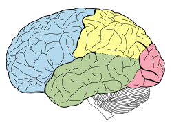
Lobes of the brain NL.svg
Lobes of the brain. Parietal lobe is yellow, and the posterior portion is near the red region.
Gray726-Brodman.svg
Lateral surface of the brain with Brodmann's areas numbered. (#5 and #7 in upper right)
Details
Identifiers
Latin cortex parietalis posterior
Anatomical terms of neuroanatomy

The posterior parietal cortex (the portion of parietal neocortex posterior to the primary somatosensory cortex) plays an important role in planned movements, spatial reasoning, and attention.

Contents

Damage to the posterior parietal cortex can produce a variety of sensorimotor deficits, including deficits in the perception and memory of spatial relationships, inaccurate reaching and grasping, in the control of eye movement, and inattention. The two most striking consequences of PPC damage are apraxia and hemispatial neglect. [1]

Anatomy

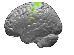
Posterior Parietal Lobe.jpg (Posterior parietal cortex (light green) is shown at the posterior area of the parietal lobe.)

The posterior parietal cortex receives input from the three sensory systems that play roles in the localization of the body and external objects in space: the visual system, the auditory system, and the somatosensory system. In turn, much of the output of the posterior parietal cortex goes to areas of frontal motor cortex: the dorsolateral prefrontal cortex, various areas of the secondary motor cortex, and the frontal eye field.

The posterior parietal cortex is divided by the intraparietal sulcus to form the dorsal superior parietal lobule and the ventral inferior parietal lobule. [2] [3] [4] Brodmann area 7 is part of the superior parietal lobule, [2] [5] but some sources include Brodmann area 5. [5] The inferior parietal lobule is further subdivided into the supramarginal gyrus, the temporoparietal junction, and the angular gyrus. [2] [3] [4] The inferior parietal lobule corresponds to Brodmann areas 39 and 40. [2] [4]

Functions

Motor

The posterior parietal cortex has been understood to have separate representations for different motor effectors (e.g. arm vs. eye). [6]

In addition to separation based on effector type, some regions are activated during both decision and execution, while other regions are only active during execution. In one study, single cell recordings showed activity in parietal reach region while non-human primates decided whether to reach or make a saccade to a target, and activity persisted during the chosen movement if and only if the monkey chose to make a reaching movement. However, cells in area 5d were only active after the decision was made to reach with the arm. [7] Another study found that neurons in area 5d only encoded the next movement in a sequence of reach movements, and not reach movements later in the sequence. [8]

In another single-cell recording experiment, neurons in parietal reach region exhibited responses consistent with either of two target locations in a sequence of planned reaching movements, suggesting that different parts of a planned sequence of locations can be represented in parallel in parietal reach region. [9]

Posterior parietal cortex appears to be involved in learning motor skills. In a PET study, researchers had subjects learn to trace a maze with their hand. Activation in right posterior parietal cortex was observed during the task, and decreased activation was associated with the number of errors made. [10] Learning a brain-computer interface produces a similar pattern: posterior parietal cortex activation decreased as subjects became more proficient. [11] One study found that novice artists have increased blood flow in the right posterior parietal compared to expert artists when challenged with art-related tasks. [12]

In a study conducted by neuroscientists at New York University, coherent patterns of firing of neurons in the brain's PPC were associated with coordination of different effectors. The researchers examined neurological activity of macaque monkeys while having them perform a variety of tasks that required them to either reach and to simultaneously employ rapid eye movements (saccades) or to only use saccades. The coherent pattern of the firing of neurons in the PPC were only seen when both the eyes and arms were required to move for the same task, but not for tasks that involved only saccades. [13]

In addition, neurons in posterior parietal cortex encode various aspects of the planned action simultaneously. Kuang and colleagues found that PPC neurons encode not only the planned physical movement, but also the anticipated visual consequence of the intended movement during the planning period. [14]

Other

Studies implicate the temporoparietal junction in exogenous or stimulus-driven attention, while the superior parietal lobule shows transient activation for self-directed switches in attention. [15] Maintaining spatial attention depends on the right posterior parietal cortex; lesions in a region between the intraparietal sulcus and inferior parietal lobule in right PPC were significantly associated with deficits in sustained spatial attention. [16]

Posterior parietal cortex is consistently activated during episodic retrieval, but most hypotheses as to why this is are speculative and usually make some connection between attention and episodic recall. [2] [3]

Damage to posterior parietal cortex results in deficits in visual working memory. [17] Patients could name objects that they had previously seen, but were impaired at recognizing previously presented objects, even if these objects had a familiar name.

In a different working memory paradigm, participants were required to make different responses to the same stimuli (letters X/Y) based on previous stimuli. [18] The previous stimuli consisted of lower-level context (letters A/B) and higher level context (numbers 1/2). The lower context specified the appropriate responses to the X/Y stimuli, while the higher level context signaled a change in the effect of the lower level context. Posterior parietal cortex was activated by lower-level context updates but not by higher-level context updates.

Posterior parietal cortex is also activated during reasoning tasks, and some of the areas activated for reasoning tend to also show activation for mathematics or calculation. [19]

There is also evidence indicating that it plays a role in perception of pain. [20]

Recent findings have suggested that feelings of "free will" at least partially originate in this area. [21] [22]

Related Research Articles

<span class="mw-page-title-main">Parietal lobe</span> Part of the brain responsible for sensory input and some language processing

The parietal lobe is one of the four major lobes of the cerebral cortex in the brain of mammals. The parietal lobe is positioned above the temporal lobe and behind the frontal lobe and central sulcus.

<span class="mw-page-title-main">Precuneus</span> Region of the parietal lobe of the brain

In neuroanatomy, the precuneus is the portion of the superior parietal lobule on the medial surface of each brain hemisphere. It is located in front of the cuneus. The precuneus is bounded in front by the marginal branch of the cingulate sulcus, at the rear by the parieto-occipital sulcus, and underneath by the subparietal sulcus. It is involved with episodic memory, visuospatial processing, reflections upon self, and aspects of consciousness.

<span class="mw-page-title-main">Motor cortex</span> Region of the cerebral cortex

The motor cortex is the region of the cerebral cortex involved in the planning, control, and execution of voluntary movements. The motor cortex is an area of the frontal lobe located in the posterior precentral gyrus immediately anterior to the central sulcus.

A gamma wave or gamma rhythm is a pattern of neural oscillation in humans with a frequency between 25 and 140 Hz, the 40 Hz point being of particular interest. Gamma rhythms are correlated with large-scale brain network activity and cognitive phenomena such as working memory, attention, and perceptual grouping, and can be increased in amplitude via meditation or neurostimulation. Altered gamma activity has been observed in many mood and cognitive disorders such as Alzheimer's disease, epilepsy, and schizophrenia.

Visual search is a type of perceptual task requiring attention that typically involves an active scan of the visual environment for a particular object or feature among other objects or features. Visual search can take place with or without eye movements. The ability to consciously locate an object or target amongst a complex array of stimuli has been extensively studied over the past 40 years. Practical examples of using visual search can be seen in everyday life, such as when one is picking out a product on a supermarket shelf, when animals are searching for food among piles of leaves, when trying to find a friend in a large crowd of people, or simply when playing visual search games such as Where's Wally?

<span class="mw-page-title-main">Premotor cortex</span>

The premotor cortex is an area of the motor cortex lying within the frontal lobe of the brain just anterior to the primary motor cortex. It occupies part of Brodmann's area 6. It has been studied mainly in primates, including monkeys and humans. The functions of the premotor cortex are diverse and not fully understood. It projects directly to the spinal cord and therefore may play a role in the direct control of behavior, with a relative emphasis on the trunk muscles of the body. It may also play a role in planning movement, in the spatial guidance of movement, in the sensory guidance of movement, in understanding the actions of others, and in using abstract rules to perform specific tasks. Different subregions of the premotor cortex have different properties and presumably emphasize different functions. Nerve signals generated in the premotor cortex cause much more complex patterns of movement than the discrete patterns generated in the primary motor cortex.

<span class="mw-page-title-main">Intraparietal sulcus</span> Sulcus on the lateral surface of the parietal lobe

The intraparietal sulcus (IPS) is located on the lateral surface of the parietal lobe, and consists of an oblique and a horizontal portion. The IPS contains a series of functionally distinct subregions that have been intensively investigated using both single cell neurophysiology in primates and human functional neuroimaging. Its principal functions are related to perceptual-motor coordination and visual attention, which allows for visually-guided pointing, grasping, and object manipulation that can produce a desired effect.

Attentional shift occurs when directing attention to a point increases the efficiency of processing of that point and includes inhibition to decrease attentional resources to unwanted or irrelevant inputs. Shifting of attention is needed to allocate attentional resources to more efficiently process information from a stimulus. Research has shown that when an object or area is attended, processing operates more efficiently. Task switching costs occur when performance on a task suffers due to the increased effort added in shifting attention. There are competing theories that attempt to explain why and how attention is shifted as well as how attention is moved through space.

<span class="mw-page-title-main">Supplementary motor area</span> Midline region in front of the motor cortex of the brain

The supplementary motor area (SMA) is a part of the motor cortex of primates that contributes to the control of movement. It is located on the midline surface of the hemisphere just in front of the primary motor cortex leg representation. In monkeys the SMA contains a rough map of the body. In humans the body map is not apparent. Neurons in the SMA project directly to the spinal cord and may play a role in the direct control of movement. Possible functions attributed to the SMA include the postural stabilization of the body, the coordination of both sides of the body such as during bimanual action, the control of movements that are internally generated rather than triggered by sensory events, and the control of sequences of movements. All of these proposed functions remain hypotheses. The precise role or roles of the SMA is not yet known.

The lateral intraparietal cortex is found in the intraparietal sulcus of the brain. This area is most likely involved in eye movement, as electrical stimulation evokes saccades of the eyes. It is also thought to contribute to working memory associated with guiding eye movement, examined using a delayed saccade task described below:

  1. A subject focuses on a fixation point at the center of a computer screen.
  2. A target is presented at a peripheral location on the screen.
  3. The target is removed and followed by a variable-length delay period.
  4. The initial focus point in the middle of the screen is removed.
  5. The subject's task is to make a saccade to the location of the target.
<span class="mw-page-title-main">Primary motor cortex</span> Brain region

The primary motor cortex is a brain region that in humans is located in the dorsal portion of the frontal lobe. It is the primary region of the motor system and works in association with other motor areas including premotor cortex, the supplementary motor area, posterior parietal cortex, and several subcortical brain regions, to plan and execute voluntary movements. Primary motor cortex is defined anatomically as the region of cortex that contains large neurons known as Betz cells, which, along with other cortical neurons, send long axons down the spinal cord to synapse onto the interneuron circuitry of the spinal cord and also directly onto the alpha motor neurons in the spinal cord which connect to the muscles.

Visual object recognition refers to the ability to identify the objects in view based on visual input. One important signature of visual object recognition is "object invariance", or the ability to identify objects across changes in the detailed context in which objects are viewed, including changes in illumination, object pose, and background context.

Michael Steven Anthony Graziano is an American scientist and novelist who is currently a professor of Psychology and Neuroscience at Princeton University. His scientific research focuses on the brain basis of awareness. He has proposed the "attention schema" theory, an explanation of how, and for what adaptive advantage, brains attribute the property of awareness to themselves. His previous work focused on how the cerebral cortex monitors the space around the body and controls movement within that space. Notably he has suggested that the classical map of the body in motor cortex, the homunculus, is not correct and is better described as a map of complex actions that make up the behavioral repertoire. His publications on this topic have had a widespread impact among neuroscientists but have also generated controversy. His novels rely partly on his background in psychology and are known for surrealism or magic realism. Graziano also composes music including symphonies and string quartets.

Richard Alan Andersen is an American neuroscientist. He is the James G. Boswell Professor of Neuroscience at the California Institute of Technology in Pasadena, California. His research focuses on visual physiology with an emphasis on translational research to humans in the field of neuroprosthetics, brain-computer interfaces, and cortical repair.

Transsaccadic memory is the neural process that allows humans to perceive their surroundings as a seamless, unified image despite rapid changes in fixation points. Transsaccadic memory is a relatively new topic of interest in the field of psychology. Conflicting views and theories have spurred several types of experiments intended to explain transsaccadic memory and the neural mechanisms involved.

Form perception is the recognition of visual elements of objects, specifically those to do with shapes, patterns and previously identified important characteristics. An object is perceived by the retina as a two-dimensional image, but the image can vary for the same object in terms of the context with which it is viewed, the apparent size of the object, the angle from which it is viewed, how illuminated it is, as well as where it resides in the field of vision. Despite the fact that each instance of observing an object leads to a unique retinal response pattern, the visual processing in the brain is capable of recognizing these experiences as analogous, allowing invariant object recognition. Visual processing occurs in a hierarchy with the lowest levels recognizing lines and contours, and slightly higher levels performing tasks such as completing boundaries and recognizing contour combinations. The highest levels integrate the perceived information to recognize an entire object. Essentially object recognition is the ability to assign labels to objects in order to categorize and identify them, thus distinguishing one object from another. During visual processing information is not created, but rather reformatted in a way that draws out the most detailed information of the stimulus.

Gain field encoding is a hypothesis about the internal storage and processing of limb motion in the brain. In the motor areas of the brain, there are neurons which collectively have the ability to store information regarding both limb positioning and velocity in relation to both the body (intrinsic) and the individual's external environment (extrinsic). The input from these neurons is taken multiplicatively, forming what is referred to as a gain field. The gain field works as a collection of internal models off of which the body can base its movements. The process of encoding and recalling these models is the basis of muscle memory.

<span class="mw-page-title-main">Ventrolateral prefrontal cortex</span> Part of the prefrontal cortex of the brain

The ventrolateral prefrontal cortex (VLPFC) is a section of the prefrontal cortex located on the inferior frontal gyrus, bounded superiorly by the inferior frontal sulcus and inferiorly by the lateral sulcus. It is attributed to the anatomical structures of Brodmann's area (BA) 47, 45 and 44.

Social cognitive neuroscience is the scientific study of the biological processes underpinning social cognition. Specifically, it uses the tools of neuroscience to study "the mental mechanisms that create, frame, regulate, and respond to our experience of the social world". Social cognitive neuroscience uses the epistemological foundations of cognitive neuroscience, and is closely related to social neuroscience. Social cognitive neuroscience employs human neuroimaging, typically using functional magnetic resonance imaging (fMRI). Human brain stimulation techniques such as transcranial magnetic stimulation and transcranial direct-current stimulation are also used. In nonhuman animals, direct electrophysiological recordings and electrical stimulation of single cells and neuronal populations are utilized for investigating lower-level social cognitive processes.

Michael E. Goldberg, also known as Mickey Goldberg, is an American neuroscientist and David Mahoney Professor at Columbia University. He is known for his work on the mechanisms of the mammalian eye in relation to brain activity. He served as president of the Society for Neuroscience from 2009 to 2010.
References

  1. Pinel, John P.J. Biopsychology Seventh Edition. Pearson Education Inc., 2009
  2. 1 2 3 4 5 Cabeza R., Ciaramelli E., Olson I. R., Moscovitch M. (2008). "The parietal cortex and episodic memory: an attentional account". Nature Reviews Neuroscience. 9 (8): 613–625. doi:10.1038/nrn2459. PMC   2692883 . PMID   18641668.{{cite journal}}: CS1 maint: multiple names: authors list (link)
  3. 1 2 3 Hutchinson J. B., Uncapher M. R., Wagner A. D. (2009). "Posterior parietal cortex and episodic retrieval: Convergent and divergent effects of attention and memory". Learning & Memory. 16 (6): 343–356. doi: 10.1101/lm.919109 . PMC   2704099 . PMID   19470649.{{cite journal}}: CS1 maint: multiple names: authors list (link)
  4. 1 2 3 Martin, R. E. (n.d.). Let’s Get to Know the Parietal Lobes! [PDF]. Retrieved from http://gablab.mit.edu/downloads/Parietal_Primer.pdf Archived 2018-02-18 at the Wayback Machine
  5. 1 2 Scheperjans F., Hermann K., Eickhoff S. B., Amunts K., Schleicher A., Zilles K. (2007). "Observer-Independent Cytoarchitectonic Mapping of the Human Superior Parietal Cortex". Cerebral Cortex. 18 (4): 846–867. doi: 10.1093/cercor/bhm116 . PMID   17644831.{{cite journal}}: CS1 maint: multiple names: authors list (link)
  6. Hwang E., Hauschild M., Wilke M., Andersen R. (2012). "Inactivation of the Parietal Reach Region Causes Optic Ataxia, Impairing Reaches but Not Saccades". Neuron. 76 (5): 1021–1029. doi: 10.1016/j.neuron.2012.10.030 . PMC   3597097 . PMID   23217749.{{cite journal}}: CS1 maint: multiple names: authors list (link)
  7. Cui H., Andersen R. A. (2011). "Different Representations of Potential and Selected Motor Plans by Distinct Parietal Areas". Journal of Neuroscience. 31 (49): 18130–18136. doi: 10.1523/jneurosci.6247-10.2011 . PMC   3327481 . PMID   22159124.
  8. Li Y., Cui H. (2013). "Dorsal Parietal Area 5 Encodes Immediate Reach in Sequential Arm Movements". Journal of Neuroscience. 33 (36): 14455–14465. doi: 10.1523/jneurosci.1162-13.2013 . PMC   6618382 . PMID   24005297.
  9. Baldauf D., Cui H., Andersen R. A. (2008). "The Posterior Parietal Cortex Encodes in Parallel Both Goals for Double-Reach Sequences". Journal of Neuroscience. 28 (40): 10081–10089. doi: 10.1523/jneurosci.3423-08.2008 . PMC   2744218 . PMID   18829966.{{cite journal}}: CS1 maint: multiple names: authors list (link)
  10. Van Mier H. I., Perlmutter J. S., Petersen S. E. (2004). "Functional Changes in Brain Activity During Acquisition and Practice of Movement Sequences". Motor Control. 8 (4): 500–520. doi:10.1123/mcj.8.4.500. PMID   15585904.{{cite journal}}: CS1 maint: multiple names: authors list (link)
  11. Wander J. D., Blakely T., Miller K. J., Weaver K. E., Johnson L. A., Olson J. D., Ojemann J. G. (2013). "Distributed cortical adaptation during learning of a brain-computer interface task". Proceedings of the National Academy of Sciences. 110 (26): 10818–10823. Bibcode:2013PNAS..11010818W. doi: 10.1073/pnas.1221127110 . PMC   3696802 . PMID   23754426.{{cite journal}}: CS1 maint: multiple names: authors list (link)
  12. Solso, Robert (February 2001). "Brain Activities in a Skilled versus a Novice Artist: An fMRI Study". Leonardo. 34 (1): 31–34. doi:10.1162/002409401300052479. S2CID   7126922.
  13. Dean H., Hagan M., Pesaran B. (2012). "Only Coherent Spiking in Posterior Parietal Cortex Coordinates Looking and Reaching". Neuron. 73 (4): 829–841. doi:10.1016/j.neuron.2011.12.035. PMC   3315591 . PMID   22365554.{{cite journal}}: CS1 maint: multiple names: authors list (link)
  14. Kuang, S.; Morel, P.; Gail, A. (2016). "Planning Movements in Visual and Physical Space in Monkey Posterior Parietal Cortex". Cerebral Cortex. 26 (2): 731–747. doi: 10.1093/cercor/bhu312 . PMID   25576535.
  15. Behrmann M., Geng J. J., Shomstein S. (2004). "Parietal cortex and attention". Current Opinion in Neurobiology. 14 (2): 212–217. doi:10.1016/j.conb.2004.03.012. PMID   15082327. S2CID   7789667.{{cite journal}}: CS1 maint: multiple names: authors list (link)
  16. Malhotra P., Coulthard E. J., Husain M. (2009). "Role of right posterior parietal cortex in maintaining attention to spatial locations over time". Brain. 132 (3): 645–660. doi: 10.1093/brain/awn350 . PMC   2664449 . PMID   19158107.{{cite journal}}: CS1 maint: multiple names: authors list (link)
  17. Berryhill M. E., Olson I. R. (2008). "Is the posterior parietal lobe involved in working memory retrieval? Evidence from patients with bilateral parietal lobe damage". Neuropsychologia. 46 (7): 1767–1774. doi:10.1016/j.neuropsychologia.2008.01.009. PMC   2441642 . PMID   18308348.
  18. Nee D. E., Brown J. W. (2012). "Dissociable Frontal-Striatal and Frontal-Parietal Networks Involved in Updating Hierarchical Contexts in Working Memory". Cerebral Cortex. 23 (9): 2146–2158. doi: 10.1093/cercor/bhs194 . PMC   3841420 . PMID   22798339.
  19. Wendelken C (2015). "Meta-analysis: how does posterior parietal cortex contribute to reasoning?". Frontiers in Human Neuroscience. 8: 1042. doi: 10.3389/fnhum.2014.01042 . PMC   4301007 . PMID   25653604.
  20. Witting N, Kupers RC, Svensson P, Arendt-Nielsen L, Gjedde A, Jensen TS (2001). "Experimental brush-evoked allodynia activates posterior parietal cortex". Neurology. 57 (10): 1817–24. doi:10.1212/wnl.57.10.1817. PMID   11723270. S2CID   8586536.{{cite journal}}: CS1 maint: multiple names: authors list (link)
  21. Desmurget M., Reilly K. T., Richard N., Szathmari A., Mottolese C., Sirigu A. (2009). "Movement Intention After Parietal Cortex Stimulation in Humans". Science. 324 (5928): 811–813. Bibcode:2009Sci...324..811D. doi:10.1126/science.1169896. PMID   19423830. S2CID   6555881.{{cite journal}}: CS1 maint: multiple names: authors list (link)
  22. Haggard P (2009). "The Sources of Human Volition". Science. 324 (5928): 731–733. Bibcode:2009Sci...324..731H. doi:10.1126/science.1173827. PMID   19423807. S2CID   206519896.