Inferior frontal gyrus

Last updated
Inferior frontal gyrus
FrontalCaptsLateral.png
Inferior frontal gyrus of the human brain, gyrus frontalis inferior.
Gray726 inferior frontal gyrus.png
Lateral surface of left hemisphere viewed from the side. Inferior frontal gyrus shown in yellow.
Details
Part of Frontal lobe
PartsPars opercularis, Pars triangularis, Pars orbitalis
Artery Middle cerebral
Identifiers
Latin gyrus frontalis inferior
NeuroNames 85
NeuroLex ID birnlex_873
TA98 A14.1.09.113
A15.2.07.058
TA2 5447
FMA 61860
Anatomical terms of neuroanatomy

The inferior frontal gyrus (IFG), (gyrus frontalis inferior), is the lowest positioned gyrus of the frontal gyri, of the frontal lobe, and is part of the prefrontal cortex.

Contents

Its superior border is the inferior frontal sulcus (which divides it from the middle frontal gyrus), its inferior border is the lateral sulcus (which divides it from the superior temporal gyrus) and its posterior border is the inferior precentral sulcus. Above it is the middle frontal gyrus, behind it is the precentral gyrus. [1]

The inferior frontal gyrus contains Broca's area, which is involved in language processing and speech production.

Structure

The inferior frontal gyrus is highly convoluted and has three cytoarchitecturally diverse regions. [2] The three subdivisions are an opercular part, a triangular part, and an orbital part. These divisions are marked by two rami arising from the lateral sulcus. [3] The ascending ramus separates the opercular and triangular parts. [4] The anterior (horizontal) ramus separates the triangular and orbital parts. [5]

Cytoarchitecturally the opercular part of the inferior frontal gyrus is known as Brodmann area 44 (BA44). The triangular part of the inferior frontal gyrus is known as Brodmann area 45 (BA45), and the orbital part of the inferior frontal gyrus is known as Brodmann area 47. The opercular part and the triangular part (BA44 and BA45) make up Broca's area.

Function

The inferior frontal gyrus has a number of functions including the processing of speech and language in Broca's area. Neural circuitry has been shown to connect different sites of stimulus to other regions of response including other subdivisions and also other frontal gyri. [2]

Language processing

The left opercular part of the inferior frontal gyrus is a part of the articulatory network involved in motor syllable programs. The articulatory network also contains the premotor cortex, and the anterior insula. These areas are interrelated but have specific functions in speech comprehension and production. The articulatory network acts mostly when the vocal tract moves to produce syllables. The pars opercularis acts indirectly through the motor cortex to control the motor aspect of speech production, and codes motor programs for this system, while the auditory cortex (via the temporoparietal junction in the lateral sulcus (Sylvian fissure) houses a series of sensory targets. Together, these areas function as a sensory-motor loop for syllable information coding.[ citation needed ]

In a study conducted comparing phonological and arithmetic processing and the involvement of different sections of the inferior frontal gyrus and angular gyrus, cortical activation for phonology, subtraction, and multiplication tasks was compared. The predetermined language-calculation network was limited to the left inferior frontal gyrus, angular gyrus, superior parietal lobule, and the horizontal portion of the intraparietal sulcus. The results were significant to support that there was a pattern of left lateralization for each of these tasks all activating the Perisylvian fissure network, with some general localized areas for phonology and arithmetic. It was supported that phonology activated the pars opercularis (BA44), and anterior angular gyrus, multiplication mainly implicated the pars triangularis (BA45), and the posterior angular gyrus. These systems are activated through similar neuronal processes but independently placed along the network.[ citation needed ]

Language comprehension and production

Most language processing takes place in Broca's area usually in the left hemisphere. [9] Damage to this region often results in a type of non-fluent aphasia known as Broca's aphasia. Broca's area is made up of the pars opercularis and the pars triangularis, both of which contribute to verbal fluency, but each has its own specific contribution. The pars opercularis (BA44) is involved in language production and phonological processing due to its connections with motor areas of the mouth and tongue. The pars triangularis (BA45) is involved in semantic processing. Characteristics of Broca's aphasia include agrammatic speech, relatively good language comprehension, poor repetition, and difficulty speaking mostly uttering short sentences made up mostly of nouns. The left IFG has also been suggested to play a role in inhibitory processes, including the tendency to inhibit learning from undesirable information. For example, transcranial magnetic stimulation to the left IFG has been shown to release such inhibition, increasing the ability to learn from undesirable information. [10]

The right opercular part of the IFG, (BA44) has been implicated in go/no go tasks. [11] In these tasks, the participant encounters a preliminary task (for instance repeatedly pressing a button), and then must halt this task whenever a "no go" signal is presented, ultimately measuring a level of impulse control through inhibition of a prepotent response. It seems that the same area is also implicated in risk aversion: a study found that higher risk aversion correlated with higher activity at IFG. [12] This might be explained as an inhibition signal to accept a risky option. Disruption of activity of this area with transcranial direct-current stimulation (tDCS) leads to change in risk attitudes, as behaviorally demonstrated by choices over risky outcomes. [13] [14]

Additional images

Related Research Articles

<span class="mw-page-title-main">Language center</span> Speech processing areas of the brain

In neuroscience and psychology, the term language center refers collectively to the areas of the brain which serve a particular function for speech processing and production. Language is a core system that gives humans the capacity to solve difficult problems and provides them with a unique type of social interaction. Language allows individuals to attribute symbols to specific concepts, and utilize them through sentences and phrases that follow proper grammatical rules. Finally, speech is the mechanism by which language is orally expressed.

<span class="mw-page-title-main">Broca's area</span> Speech production region in the dominant hemisphere of the hominid brain

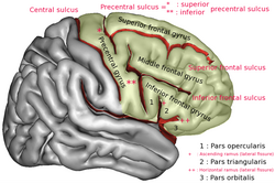
Broca's area, or the Broca area, is a region in the frontal lobe of the dominant hemisphere, usually the left, of the brain with functions linked to speech production.

Articles related to anatomy include:

<span class="mw-page-title-main">Cingulate cortex</span> Part of the brain within the cerebral cortex

The cingulate cortex is a part of the brain situated in the medial aspect of the cerebral cortex. The cingulate cortex includes the entire cingulate gyrus, which lies immediately above the corpus callosum, and the continuation of this in the cingulate sulcus. The cingulate cortex is usually considered part of the limbic lobe.

<span class="mw-page-title-main">Brodmann area</span> Region of the brain

A Brodmann area is a region of the cerebral cortex, in the human or other primate brain, defined by its cytoarchitecture, or histological structure and organization of cells. The concept was first introduced by the German anatomist Korbinian Brodmann in the early 20th century. Brodmann mapped the human brain based on the varied cellular structure across the cortex and identified 52 distinct regions, which he numbered 1 to 52. These regions, or Brodmann areas, correspond with diverse functions including sensation, motor control, and cognition.

<span class="mw-page-title-main">Frontal lobe</span> Part of the brain

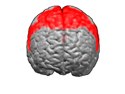
The frontal lobe is the largest of the four major lobes of the brain in mammals, and is located at the front of each cerebral hemisphere. It is parted from the parietal lobe by a groove between tissues called the central sulcus and from the temporal lobe by a deeper groove called the lateral sulcus. The most anterior rounded part of the frontal lobe is known as the frontal pole, one of the three poles of the cerebrum.

<span class="mw-page-title-main">Brodmann area 6</span>

Brodmann area 6 (BA6) is part of the frontal cortex in the human brain. Situated just anterior to the primary motor cortex (BA4), it is composed of the premotor cortex and, medially, the supplementary motor area (SMA). This large area of the frontal cortex is believed to play a role in planning complex, coordinated movements.

<span class="mw-page-title-main">Brodmann area 44</span> Brain area

Brodmann area 44, or BA44, is part of the frontal cortex in the human brain. Situated just anterior to premotor cortex (BA6) and on the lateral surface, inferior to BA9.

<span class="mw-page-title-main">Brodmann area 45</span> Brain area

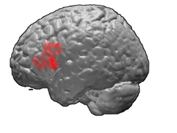
Brodmann area 45 (BA45), is part of the frontal cortex in the human brain. It is situated on the lateral surface, inferior to BA9 and adjacent to BA46.
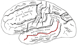
Expressive language disorder is one of the "specific developmental disorders of speech and language" recognised by the tenth edition of the International Classification of Diseases (ICD-10). As of the eleventh edition, it is considered to be covered by the various categories of developmental language disorder. Transition to the ICD-11 will take place at a different time in different countries.

<span class="mw-page-title-main">Language processing in the brain</span> How humans use words to communicate

In psycholinguistics, language processing refers to the way humans use words to communicate ideas and feelings, and how such communications are processed and understood. Language processing is considered to be a uniquely human ability that is not produced with the same grammatical understanding or systematicity in even human's closest primate relatives.

<span class="mw-page-title-main">Gyrus</span> Ridge on the cerebral cortex of the brain

In neuroanatomy, a gyrus is a ridge on the cerebral cortex. It is generally surrounded by one or more sulci. Gyri and sulci create the folded appearance of the brain in humans and other mammals.

<span class="mw-page-title-main">Middle cerebral artery</span> Paired artery that supplies blood to the cerebrum

The middle cerebral artery (MCA) is one of the three major paired cerebral arteries that supply blood to the cerebrum. The MCA arises from the internal carotid artery and continues into the lateral sulcus where it then branches and projects to many parts of the lateral cerebral cortex. It also supplies blood to the anterior temporal lobes and the insular cortices.

<span class="mw-page-title-main">Lobes of the brain</span> Parts of the cerebrum

The lobes of the brain are the major identifiable zones of the human cerebral cortex, and they comprise the surface of each hemisphere of the cerebrum. The two hemispheres are roughly symmetrical in structure, and are connected by the corpus callosum. They traditionally have been divided into four lobes, but are today considered as having six lobes each. The lobes are large areas that are anatomically distinguishable, and are also functionally distinct to some degree. Each lobe of the brain has numerous ridges, or gyri, and furrows, the sulci that constitute further subzones of the cortex. The expression "lobes of the brain" usually refers only to those of the cerebrum, not to the distinct areas of the cerebellum.

<span class="mw-page-title-main">Olfactory tract</span> Part of the olfactory system

The olfactory tract is a bilateral bundle of afferent nerve fibers from the mitral and tufted cells of the olfactory bulb that connects to several target regions in the brain, including the piriform cortex, amygdala, and entorhinal cortex. It is a narrow white band, triangular on coronal section, the apex being directed upward.

<span class="mw-page-title-main">Foix–Chavany–Marie syndrome</span> Medical condition

Foix–Chavany–Marie syndrome (FCMS), also known as bilateral opercular syndrome, is a neuropathological disorder characterized by paralysis of the facial, tongue, pharynx, and masticatory muscles of the mouth that aid in chewing. The disorder is primarily caused by thrombotic and embolic strokes, which cause a deficiency of oxygen in the brain. As a result, bilateral lesions may form in the junctions between the frontal lobe and temporal lobe, the parietal lobe and cortical lobe, or the subcortical region of the brain. FCMS may also arise from defects existing at birth that may be inherited or nonhereditary. Symptoms of FCMS can be present in a person of any age and it is diagnosed using automatic-voluntary dissociation assessment, psycholinguistic testing, neuropsychological testing, and brain scanning. Treatment for FCMS depends on the onset, as well as on the severity of symptoms, and it involves a multidisciplinary approach.

<span class="mw-page-title-main">Superior longitudinal fasciculus</span> Association fiber tract of the brain

The superior longitudinal fasciculus (SLF) is an association tract in the brain that is composed of three separate components. It is present in both hemispheres and can be found lateral to the centrum semiovale and connects the frontal, occipital, parietal, and temporal lobes. This bundle of tracts (fasciculus) passes from the frontal lobe through the operculum to the posterior end of the lateral sulcus where they either radiate to and synapse on neurons in the occipital lobe, or turn downward and forward around the putamen and then radiate to and synapse on neurons in anterior portions of the temporal lobe.

<span class="mw-page-title-main">Superior temporal sulcus</span> Part of the brains temporal lobe

In the human brain, the superior temporal sulcus (STS) is the sulcus separating the superior temporal gyrus from the middle temporal gyrus in the temporal lobe of the brain. A sulcus is a deep groove that curves into the largest part of the brain, the cerebrum, and a gyrus is a ridge that curves outward of the cerebrum.

<span class="mw-page-title-main">Orbital part of inferior frontal gyrus</span>

The orbital part of inferior frontal gyrus also known as the pars orbitalis is the orbital part of the inferior frontal gyrus.

References

  1. Nolte (2002), The Human Brain, ISBN   978-0-323-01320-8 photos on p526 & p.546
  2. 1 2 Greenlee, JD; et al. (1 August 2007). "Functional connections within the human inferior frontal gyrus". The Journal of Comparative Neurology. 503 (4): 550–9. doi:10.1002/cne.21405. PMID   17534935. S2CID   5685566.
  3. "anterior ramus of lateral cerebral sulcus". TheFreeDictionary.com.
  4. Gaillard, Frank. "Ascending ramus of the lateral sulcus | Radiology Reference Article | Radiopaedia.org". Radiopaedia.
  5. Gaillard, Frank. "Anterior ramus of the lateral sulcus | Radiology Reference Article | Radiopaedia.org". Radiopaedia.
  6. Schremm, A; et al. (January 2018). "Cortical thickness of planum temporale and pars opercularis in native language tone processing". Brain and Language. 176: 42–47. doi: 10.1016/j.bandl.2017.12.001 . PMID   29223785.
  7. Tyler, LK; et al. (2005). "Temporal and frontal systems in speech comprehension: An fMRI study of past tense processing". Neuropsychologia. 43 (13): 1963–1974. doi:10.1016/j.neuropsychologia.2005.03.008. PMID   16168736. S2CID   16112201.
  8. Elmer, Stefan (29 November 2016). "Broca Pars Triangularis Constitutes a "Hub" of the Language-Control Network during Simultaneous Language Translation". Frontiers in Human Neuroscience. 10: 491. doi: 10.3389/fnhum.2016.00491 . PMC   5040713 . PMID   27746729.
  9. The "dominant inferior frontal convolution" —Fauci, et.al, eds. (1998), Harrison's Principles of Internal Medicine, 14th Edition, Companion Handbook, ISBN   978-0-07-021530-6 {{citation}}: |first= has generic name (help)CS1 maint: multiple names: authors list (link). p.1055
  10. Sharot, T., Kanai, R., Marston, D., Korn, C. W., Rees, G. & Dolan, R.J. (2012) Selectively Altering Belief Formation in the Human Brain. Proceedings of the National Academy of Sciences, 109 (42), 17058–17062.
  11. Aron AR, Robbins TW, Poldrack RA (2004). "Inhibition and the right inferior frontal cortex". Trends Cogn Sci. 8 (4): 170–177. doi:10.1016/j.tics.2004.02.010. PMID   15050513. S2CID   19332756.
  12. Christopoulos, GI.; Tobler, PN.; Bossaerts, P.; Dolan, RJ.; Schultz, W. (Oct 2009). "Neural correlates of value, risk, and risk aversion contributing to decision making under risk". J Neurosci. 29 (40): 12574–83. doi:10.1523/JNEUROSCI.2614-09.2009. PMC   2794196 . PMID   19812332.
  13. Knoch D, Gianotti LR, Pascual-Leone A, Treyer V, Regard M, Hohmann M, Brugger P (2006). "Disruption of right prefrontal cortex by low-frequency repetitive transcranial magnetic stimulation induces risk-taking behavior" (PDF). J Neurosci. 26 (24): 6469–6472. doi: 10.1523/JNEUROSCI.0804-06.2006 . PMC   6674035 . PMID   16775134.
  14. Fecteau S, Pascual-Leone A, Zald DH, Liguori P, Théoret H, Boggio PS, Fregni F (2007). "Activation of prefrontal cortex by transcranial direct current stimulation reduces appetite for risk during ambiguous decision making". J Neurosci. 27 (23): 6212–6218. doi: 10.1523/JNEUROSCI.0314-07.2007 . PMC   6672163 . PMID   17553993.