
A Brodmann area is a region of the cerebral cortex, in the human or other primate brain, defined by its cytoarchitecture, or histological structure and organization of cells. The concept was first introduced by the German anatomist Korbinian Brodmann in the early 20th century. Brodmann mapped the human brain based on the varied cellular structure across the cortex and identified 52 distinct regions, which he numbered 1 to 52. These regions, or Brodmann areas, correspond with diverse functions including sensation, motor control, and cognition.

Brodmann area 23 (BA23) is a region in the brain that lies inside the posterior cingulate cortex. It lies between Brodmann area 30 and Brodmann area 31 and is located on the medial wall of the cingulate gyrus between the callosal sulcus and the cingulate sulcus.
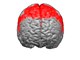
Brodmann area 6 (BA6) is part of the frontal cortex in the human brain. Situated just anterior to the primary motor cortex (BA4), it is composed of the premotor cortex and, medially, the supplementary motor area (SMA). This large area of the frontal cortex is believed to play a role in planning complex, coordinated movements.

Brodmann area 10 is the anterior-most portion of the prefrontal cortex in the human brain. BA10 was originally defined broadly in terms of its cytoarchitectonic traits as they were observed in the brains of cadavers, but because modern functional imaging cannot precisely identify these boundaries, the terms anterior prefrontal cortex, rostral prefrontal cortex and frontopolar prefrontal cortex are used to refer to the area in the most anterior part of the frontal cortex that approximately covers BA10—simply to emphasize the fact that BA10 does not include all parts of the prefrontal cortex.

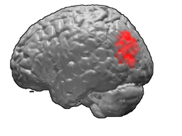
Brodmann area 44, or BA44, is part of the frontal cortex in the human brain. Situated just anterior to premotor cortex (BA6) and on the lateral surface, inferior to BA9.

Brodmann area 19, or BA 19, is part of the occipital lobe cortex in the human brain. Along with area 18, it comprises the extrastriate cortex. In humans with normal sight, extrastriate cortex is a visual association area, with feature-extracting, shape recognition, attentional, and multimodal integrating functions.

Brodmann area 20, or BA20, is part of the temporal cortex in the human brain. The region encompasses most of the ventral temporal cortex, a region believed to play a part in high-level visual processing and recognition memory.

Brodmann area 21, or BA21, is part of the temporal cortex in the human brain. The region encompasses most of the lateral temporal cortex and is also known as middle temporal area 21. In the human it corresponds approximately to the middle temporal gyrus.

Brodmann area 38, also BA38 or temporopolar area 38 (H), is part of the temporal cortex in the human brain. BA 38 is at the anterior end of the temporal lobe, known as the temporal pole.
Brodmann area 39, or BA39, is part of the parietal cortex in the human brain. BA39 encompasses the angular gyrus, lying near to the junction of temporal, occipital and parietal lobes.

Brodmann area 40 (BA40) is part of the parietal cortex in the human brain. The inferior part of BA40 is in the area of the supramarginal gyrus, which lies at the posterior end of the lateral fissure, in the inferior lateral part of the parietal lobe.

Brodmann area 11 is one of Brodmann's cytologically defined regions of the brain. It is in the orbitofrontal cortex which is above the eye sockets (orbitae). It is involved in decision making, processing rewards, and encoding new information into long-term memory.

The fusiform gyrus, also known as the lateral occipitotemporal gyrus,is part of the temporal lobe and occipital lobe in Brodmann area 37. The fusiform gyrus is located between the lingual gyrus and parahippocampal gyrus above, and the inferior temporal gyrus below. Though the functionality of the fusiform gyrus is not fully understood, it has been linked with various neural pathways related to recognition. Additionally, it has been linked to various neurological phenomena such as synesthesia, dyslexia, and prosopagnosia.

The inferior frontal gyrus (IFG),, is the lowest positioned gyrus of the frontal gyri, of the frontal lobe, and is part of the prefrontal cortex.

Brodmann area 4 refers to the primary motor cortex of the human brain. It is located in the posterior portion of the frontal lobe.

Brodmann area 22 is a Brodmann's area that is cytoarchitecturally located in the posterior superior temporal gyrus of the brain. In the left cerebral hemisphere, it is one portion of Wernicke's area. The left hemisphere BA22 helps with generation and understanding of individual words. On the right side of the brain, BA22 helps to discriminate pitch and sound intensity, both of which are necessary to perceive melody and prosody. Wernicke's area is active in processing language and consists of the left Brodmann area 22 and Brodmann area 40, the supramarginal gyrus.

Brodmann area 35, together with Brodmann area 36, comprise the perirhinal cortex. They are cytoarchitecturally defined temporal regions of the cerebral cortex.

The inferior temporal gyrus is one of three gyri of the temporal lobe and is located below the middle temporal gyrus, connected behind with the inferior occipital gyrus; it also extends around the infero-lateral border on to the inferior surface of the temporal lobe, where it is limited by the inferior sulcus. This region is one of the higher levels of the ventral stream of visual processing, associated with the representation of objects, places, faces, and colors. It may also be involved in face perception, and in the recognition of numbers and words.

