Superior temporal sulcus

Last updated
Superior temporal sulcus
250px-Gray726-STS.png
Superior temporal sulcus.png
Details
Part of Temporal lobe
Identifiers
Latin sulcus temporalis superior
NeuroNames 129
TA98 A14.1.09.145
TA2 5494
FMA 83783
Anatomical terms of neuroanatomy

In the human brain, the superior temporal sulcus (STS) is the sulcus separating the superior temporal gyrus from the middle temporal gyrus in the temporal lobe of the brain. A sulcus (plural sulci) is a deep groove that curves into the largest part of the brain, the cerebrum, and a gyrus (plural gyri) is a ridge that curves outward of the cerebrum. [1]

Contents

The STS is located under the lateral fissure, which is the fissure that separates the temporal lobe, parietal lobe, and frontal lobe. [1] The STS has an asymmetric structure between the left and right hemisphere, with the STS being longer in the left hemisphere, but deeper in the right hemisphere. [2] This asymmetrical structural organization between hemispheres has only been found to occur in the STS of the human brain. [2]

The STS has been shown to produce strong responses when subjects perceive stimuli in research areas that include theory of mind, biological motion, faces, voices, and language. [3] [4]

Language processing

Spoken language processing

The superior temporal sulcus also activates when hearing human voices. [5] It is thought to be a source of sensory encoding linked to motor output through the superior parietal-temporal areas of the brain inferred from the time course of activation. The conclusion of pertinence to vocal processing can be drawn from data showing that the regions of the STS are more active when people are listening to vocal sounds rather than non-vocal environmentally based sounds and corresponding control sounds, which can be scrambled or modulated voices. [6] These experimental results indicate the involvement of the STS in the areas of speech and language recognition.

The majority of studies find it is the middle to the posterior portion of the STS that is involved in phonological processing, with bilateral activation indicated though including a mild left hemisphere bias due to greater observed activation. However, the role of the anterior STS in the ventral pathway of speech comprehension and production has not been ruled out. [7] Evidence for the involvement of the middle portion of the STS in phonological processing comes from repetition-suppression studies, which use fMRI to pinpoint areas of the brain responsible for specialized stimulus involvement by habituating the brain to the stimulus and recording differences in stimulation response. The resulting pattern showed expected results in the middle portion of the STS. [8]

Studies using fMRI analysis to measure superior temporal sulcus activation have found that phonemes, words, sentences, and phonological cues all lead to increased activation throughout a posterior-anterior axis in the temporal lobe. [2] This pattern of activation, which most frequently occurs in the left hemisphere, has been termed the ventral stream of speech perception. [7] Many studies consistently indicate that the superior temporal sulcus activation is associated with the interpretation of phonological signals. [2] Although present research suggest that the left hemisphere of the superior temporal sulcus and its associated left ventral stream plays a role in phonological processing, the right hemisphere of the superior temporal sulcus has been connected to the perception of voice and the prosody of speech. [9]

According to the audiological pathway model supplied by Hickok and Poeppel, after the spectrotemporal analysis conducted by the auditory cortex, the STS is responsible for interpretation of vocal input through the phonological network. This implication is shown in the activation of the region in tasks of speech perception and processing, which necessarily involves access to and continuance of phonological information. By manipulating the interactions of phonological data, represented by the provision of words with high or low neighborhood density (words associated with many or few other words), the fluctuation of activity of the STS region can be seen. This changing activation links the STS with the phonological pathway. [7]

Sign language processing

Research shows that the Broca's area of the brain is activated during sign language production and processing. [10] However, while the Broca's area plays a significant role, there are additional regions such as the posterior superior temporal gyrus and the left inferior parietal lobe that also play vital roles in the processing of sign language. Therefore, sign language engages with several regions of the brain, not simply the Brocas's area. [11]

Although Broca's area is found in the frontal lobe, it receives connection from the superior temporal gyrus, including the STS. [10] Native signers are people who learned and have been using sign language, such as American Sign Language (ASL), from birth, and/or use it as their first language. [12] They often learn sign language from their parents and continue its use throughout their lifetime. [12] Sign language activates language regions of the brain, including the STS. [13] There have been studies that show activation of the STS while deaf and hearing native signers perceive sign language, suggesting the STS is tied to the linguistic processing aspect of sign language. [14] [15] It is also important to highlight the importance of the superior temporal sulcus in its involvement throughout different parts of auditory and visual processing. The superior temporal sulcus is activated during the perception of sign language - this could potentially be related to visual-spatial and linguistic processing. [16] [17]

Studies also show that there is greater activation of the middle STS in both deaf and hearing signers who acquired ASL earlier than those who acquired it later. [18]

Social processing

Studies reveal multiple social processing capabilities. [19] Research has documented activation in the STS as a result of five specific social inputs, and thus the STS is assumed to be implicated in social perception. It showed increased activation related to: theory of mind (false belief stories versus false physical stories), voices versus environmental sounds, stories versus nonsense speech, moving faces versus moving objects, and biological motion. [20] [3] It is involved in the perception of where others are gazing (joint attention) and is important in determining where others' emotions are being directed. [21]

Theory of mind

Neuroimaging studies examining the theory of mind, otherwise known as the ability to attribute mental states to others, have identified the posterior superior temporal sulcus of the right hemisphere as being involved in its processing. [2] Activation of this region in the theory of mind has been found to be best predicted by independent ratings from other groups of participants, or more specifically, how much each item in the study made them consider the protagonist's point of view. [22] Reports noted in other studies suggest a number of inconsistencies with the localization of theory of mind processing, such as the middle and anterior portions of the superior temporal sulcus having increased activation in response to theory of mind tasks. [3] Thus, further research is needed to expand upon the precise functional role of the superior temporal sulcus in the perception of theory of mind.

Face perception

A recent study identified a region of the posterior superior temporal sulcus that is preferentially activated in the interpretation of facial expressions. [23] Similarly, another study found that transcranial magnetic stimulation disrupted the neural response to faces, but not the neural response to bodies or objects. [24] The patterns of activations found in this study suggests that facial information is processed by projections in the right hemisphere from the posterior superior temporal sulcus, through the anterior superior temporal sulcus, and into the amygdala. [24] Another study showed that the resting-state functional connectivity between the right posterior superior temporal sulcus, the right occipital face area, early visual cortex, and bilateral superior temporal sulcus was positively correlated with each subject's ability to recognize facial expression. [25]

Face-voice audiovisual integration

Many studies have suggested that the posterior superior temporal sulcus is associated with the crossmodal binding of auditory and visual stimuli. [2] The activation of this posterior portion of the superior temporal sulcus was reported in the detection of audio-visual incongruences and in voice perception. [2] The posterior superior temporal sulcus has also been shown to be preferentially activated by lip reading. [26] An area of the right posterior superior temporal sulcus was characterized by a recent study by a stronger response to audiovisual stimuli compared to that of auditory or visual stimuli alone. [27] This study also identified this same region to preferentially activated in the processing of stimuli associated with people, such as faces and voices. [27] Another fMRI study found that the neural representations of audiovisual integration, non-verbal emotional signals, voice sensitivity, and face sensitivity are all localized to separate regions of the superior temporal sulcus. [28] Likewise, this study also noted that the area most sensitive to voice is located in the trunk section of the superior temporal sulcus, the area most sensitive to facial expressions is located in the posterior terminal ascending branch, and audiovisual integration of emotional signals occurs in regions that overlap with face and voice recognition areas at the bifurcation of the superior temporal sulcus. [28]

Biological motion

The superior temporal sulcus has been found to have a unique sensitivity to observable manifestations of movement understanding, which suggest that the superior temporal sulcus is heavily involved in the recognition of movements and gestures required for normal social information processing in humans. [2] In fMRI studies evaluating the interpretation of a point-light display that represents a moving human figure as a pattern of dots, a cluster of significant brain activity was observed in the posterior superior temporal sulcus of the right hemisphere in subjects that correctly identified the biological motion being shown in the point-light display. [29] Furthermore, movement perception and movement interpretation is suggested to be localized in different regions of the superior temporal sulcus, with movement perception being processed in a posterior region of the superior temporal sulcus and movement understanding being processed in a more anterior region. [29]

Neurological disorders

In studies on dysfunctional social cognition in neurological disorders, such as what is observed in people with high-functioning autism, the role of the superior temporal sulcus in processing social information has been identified as the mechanism that lies at the root of these impairments in social interpretation. [30]

Autism and schizophrenia

Children with high-functioning autism have been reported to have no significant change in superior temporal sulcus activation for biological motion compared to non-biological motion, which suggests that the superior temporal sulcus is not specifically activated in the processing of biological motion like it is in children without autism. [30] In subjects with schizophrenia, another neurological disorder associated with significant impairments to social cognition, these social impairments have been linked to an alteration in posterior superior temporal sulcus activation in affective theory of mind, emotional recognition, and the interpretation of neutral facial expressions. [31] More specifically, it was determined that schizophrenic subjects exhibited hyperactivity within the posterior superior temporal sulcus of the right hemisphere in processing neutral facial expressions, but they also exhibited hypoactivity within this same region for emotional recognition and affective theory of mind. [31] This same study also found impaired connectivity between the right and left hemispheres of the posterior superior temporal sulcus in the processing of affective theory of mind. [31] Another recent study showed an inverse relationship between glutamate concentrations within the superior temporal sulcus and neuroticism scores assessed by questionnaire was found in subjects with schizophrenia, which suggests that elevations in glutamate concentrations may act as a compensatory mechanism that allows those with schizophrenia to prevent neuroticism. [32]

Agnosia

Various disorders of the STS have been documented in which patients fail to recognize a certain stimulus, but still exhibit subcortical processing of the stimulus, this is known as an agnosia. Furthermore, agnosia is often linked to experiencing hardship in regard to stimuli recognition despite presenting otherwise normal or intact sensory functioning. Agnosia is found to disturb higher-order centers of the brain which also include cortical regions such as the posterior parietal cortex and occipitotemporal regions. [33] [34]

Pure auditory agnosia (agnosia without aphasia) is found in patients who can't identify non-speech sounds such as coughing, whistling, and crying but have no deficit in speech comprehension. Speech agnosia is known as an incapability to comprehend spoken words despite intact hearing, speech production, and reading ability. Patients show a recognition of the familiarity of a word, but are not able to recall its meaning. Phonagnosia is characterized as an inability to recognize familiar voices, while having other auditory abilities. Patients exhibited a double dissociation with either an inability to match names or faces with a certain famous voice, or to discriminate familiar voices from unfamiliar ones. Visual agnosia can be broken into separate disorders in regard to what is being recognized. [35] An inability to recognize written words is known as alexia or word blindness, while an inability to recognize familiar faces is known as prosopagnosia. Prosopagnosia has been shown to have a similar double dissociation as phonagnosia in that some patients show an impairment of memory for familiar faces while others show impairment when discriminating familiar faces from unfamiliar ones.

Dual Pathway Model:

Gregory Hickok and David Poeppel's model proposed what is known as the dual pathway or the dual-stream model. This model explores the perception and experience of speech stimuli. The model implies that two streams process information at the perception of speech - the ventral and dorsal stream. The ventral stream aids in the comprehension and recognition of speech input passing through the ears and entering the brain. On the other hand, the dorsal stream allows an individual to respond to said input as the speech stimuli further undergoes processing by the superior temporal gyrus. This correlates to the superior temporal sulcus because the dual pathway model occurs next after “spectrotemporal analysis” is carried out through the auditory cortex. [36] [37]

Determination of Speech versus Non-Speech:

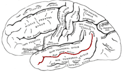
The superior temporal sulcus has an important part in the processing of human speech - specifically comprehension and perception of human voices/spoken language. According to “The superior temporal sulcus” (Howard 2023), research studies have been conducted by Blinder (2000) and Beline (2000) that examine the way in which the superior temporal sulcus reacts to various forms of stimuli especially speech and non-speech stimuli. Results show that the superior temporal sulcus is favorable towards response to human voices. [36] [38]

Phonological Neighborhoods:

Phonological neighborhoods are “neighborhoods” or groups of words that all have common or similar sounds. Research shows how much the superior temporal sulcus plays a vital role in processing and understanding of phonological neighborhoods. Words experience classification based on the amount of other words with similar sounds. High neighborhood density words describe words that are similar phonetically to several other words. On the other hand, low neighborhood density describes words that have few similar sounding words. [39] [40]

Related Research Articles

<span class="mw-page-title-main">Language center</span> Speech processing areas of the brain

In neuroscience and psychology, the term language center refers collectively to the areas of the brain which serve a particular function for speech processing and production. Language is a core system that gives humans the capacity to solve difficult problems and provides them with a unique type of social interaction. Language allows individuals to attribute symbols to specific concepts, and utilize them through sentences and phrases that follow proper grammatical rules. Finally, speech is the mechanism by which language is orally expressed.

<span class="mw-page-title-main">Broca's area</span> Speech production region in the dominant hemisphere of the hominid brain

Broca's area, or the Broca area, is a region in the frontal lobe of the dominant hemisphere, usually the left, of the brain with functions linked to speech production.

<span class="mw-page-title-main">Agnosia</span> Inability to process sensory information

Agnosia is a neurological disorder characterized by an inability to process sensory information. Often there is a loss of ability to recognize objects, persons, sounds, shapes, or smells while the specific sense is not defective nor is there any significant memory loss. It is usually associated with brain injury or neurological illness, particularly after damage to the occipitotemporal border, which is part of the ventral stream. Agnosia only affects a single modality, such as vision or hearing. More recently, a top-down interruption is considered to cause the disturbance of handling perceptual information.

<span class="mw-page-title-main">Temporal lobe</span> One of the four lobes of the mammalian brain

The temporal lobe is one of the four major lobes of the cerebral cortex in the brain of mammals. The temporal lobe is located beneath the lateral fissure on both cerebral hemispheres of the mammalian brain.

<span class="mw-page-title-main">Wernicke's area</span> Speech comprehension region in the dominant hemisphere of the hominid brain

Wernicke's area, also called Wernicke's speech area, is one of the two parts of the cerebral cortex that are linked to speech, the other being Broca's area. It is involved in the comprehension of written and spoken language, in contrast to Broca's area, which is primarily involved in the production of language. It is traditionally thought to reside in Brodmann area 22, which is located in the superior temporal gyrus in the dominant cerebral hemisphere, which is the left hemisphere in about 95% of right-handed individuals and 70% of left-handed individuals.

<span class="mw-page-title-main">Inferior frontal gyrus</span> Part of the brains prefrontal cortex

The inferior frontal gyrus (IFG),, is the lowest positioned gyrus of the frontal gyri, of the frontal lobe, and is part of the prefrontal cortex.

<span class="mw-page-title-main">Transverse temporal gyrus</span> Gyrus of the primary auditory cortex of the brain

The transverse temporal gyri, also called Heschl's gyri or Heschl's convolutions, are gyri found in the area of primary auditory cortex buried within the lateral sulcus of the human brain, occupying Brodmann areas 41 and 42. Transverse temporal gyri are superior to and separated from the planum temporale by Heschl's sulcus. Transverse temporal gyri are found in varying numbers in both the right and left hemispheres of the brain and one study found that this number is not related to the hemisphere or dominance of hemisphere studied in subjects. Transverse temporal gyri can be viewed in the sagittal plane as either an omega shape or a heart shape.

<span class="mw-page-title-main">Superior temporal gyrus</span> One of three gyri of the temporal lobe of the brain

The superior temporal gyrus (STG) is one of three gyri in the temporal lobe of the human brain, which is located laterally to the head, situated somewhat above the external ear.

<span class="mw-page-title-main">Angular gyrus</span> Gyrus of the parietal lobe of the brain

The angular gyrus is a region of the brain lying mainly in the posteroinferior region of the parietal lobe, occupying the posterior part of the inferior parietal lobule. It represents the Brodmann area 39.

<span class="mw-page-title-main">Language processing in the brain</span> How humans use words to communicate

In psycholinguistics, language processing refers to the way humans use words to communicate ideas and feelings, and how such communications are processed and understood. Language processing is considered to be a uniquely human ability that is not produced with the same grammatical understanding or systematicity in even human's closest primate relatives.

<span class="mw-page-title-main">Brodmann area 22</span>

Brodmann area 22 is a Brodmann's area that is cytoarchitecturally located in the posterior superior temporal gyrus of the brain. In the left cerebral hemisphere, it is one portion of Wernicke's area. The left hemisphere BA22 helps with generation and understanding of individual words. On the right side of the brain, BA22 helps to discriminate pitch and sound intensity, both of which are necessary to perceive melody and prosody. Wernicke's area is active in processing language and consists of the left Brodmann area 22 and Brodmann area 40, the supramarginal gyrus.

The two-streams hypothesis is a model of the neural processing of vision as well as hearing. The hypothesis, given its initial characterisation in a paper by David Milner and Melvyn A. Goodale in 1992, argues that humans possess two distinct visual systems. Recently there seems to be evidence of two distinct auditory systems as well. As visual information exits the occipital lobe, and as sound leaves the phonological network, it follows two main pathways, or "streams". The ventral stream leads to the temporal lobe, which is involved with object and visual identification and recognition. The dorsal stream leads to the parietal lobe, which is involved with processing the object's spatial location relative to the viewer and with speech repetition.

Auditory verbal agnosia (AVA), also known as pure word deafness, is the inability to comprehend speech. Individuals with this disorder lose the ability to understand language, repeat words, and write from dictation. Some patients with AVA describe hearing spoken language as meaningless noise, often as though the person speaking was doing so in a foreign language. However, spontaneous speaking, reading, and writing are preserved. The maintenance of the ability to process non-speech auditory information, including music, also remains relatively more intact than spoken language comprehension. Individuals who exhibit pure word deafness are also still able to recognize non-verbal sounds. The ability to interpret language via lip reading, hand gestures, and context clues is preserved as well. Sometimes, this agnosia is preceded by cortical deafness; however, this is not always the case. Researchers have documented that in most patients exhibiting auditory verbal agnosia, the discrimination of consonants is more difficult than that of vowels, but as with most neurological disorders, there is variation among patients.

<span class="mw-page-title-main">Middle temporal gyrus</span> One of three gyri of the temporal lobe of the brain

Middle temporal gyrus is a gyrus in the brain on the temporal lobe. It is located between the superior temporal gyrus and inferior temporal gyrus. It corresponds largely to Brodmann area 21.

<span class="mw-page-title-main">Inferior parietal lobule</span> Portion of the parietal lobe of the brain

The inferior parietal lobule lies below the horizontal portion of the intraparietal sulcus, and behind the lower part of the postcentral sulcus. Also known as Geschwind's territory after Norman Geschwind, an American neurologist, who in the early 1960s recognised its importance. It is a part of the parietal lobe.

<span class="mw-page-title-main">Lingual gyrus</span> Gyrus of the occipital lobe of the brain

The lingual gyrus, also known as the medialoccipitotemporal gyrus, is a brain structure that is linked to processing vision, especially related to letters. It is thought to also play a role in analysis of logical conditions and encoding visual memories. It is named after its shape, which is somewhat similar to a tongue. Contrary to the name, the region has little to do with speech.

Auditory agnosia is a form of agnosia that manifests itself primarily in the inability to recognize or differentiate between sounds. It is not a defect of the ear or "hearing", but rather a neurological inability of the brain to process sound meaning. While auditory agnosia impairs the understanding of sounds, other abilities such as reading, writing, and speaking are not hindered. It is caused by bilateral damage to the anterior superior temporal gyrus, which is part of the auditory pathway responsible for sound recognition, the auditory "what" pathway.

The neuroscience of music is the scientific study of brain-based mechanisms involved in the cognitive processes underlying music. These behaviours include music listening, performing, composing, reading, writing, and ancillary activities. It also is increasingly concerned with the brain basis for musical aesthetics and musical emotion. Scientists working in this field may have training in cognitive neuroscience, neurology, neuroanatomy, psychology, music theory, computer science, and other relevant fields.

<span class="mw-page-title-main">Sign language in the brain</span>

Sign language refers to any natural language which uses visual gestures produced by the hands and body language to express meaning. The brain's left side is the dominant side utilized for producing and understanding sign language, just as it is for speech. In 1861, Paul Broca studied patients with the ability to understand spoken languages but the inability to produce them. The damaged area was named Broca's area, and located in the left hemisphere’s inferior frontal gyrus. Soon after, in 1874, Carl Wernicke studied patients with the reverse deficits: patients could produce spoken language, but could not comprehend it. The damaged area was named Wernicke's area, and is located in the left hemisphere’s posterior superior temporal gyrus.

Interindividual differences in perception describes the effect that differences in brain structure or factors such as culture, upbringing and environment have on the perception of humans. Interindividual variability is usually regarded as a source of noise for research. However, in recent years, it has become an interesting source to study sensory mechanisms and understand human behavior. With the help of modern neuroimaging methods such as fMRI and EEG, individual differences in perception could be related to the underlying brain mechanisms. This has helped to explain differences in behavior and cognition across the population. Common methods include studying the perception of illusions, as they can effectively demonstrate how different aspects such as culture, genetics and the environment can influence human behavior.

References

  1. 1 2 Bui, Toai; M Das, Joe (2020), "Neuroanatomy, Cerebral Hemisphere", StatPearls, Treasure Island (FL): StatPearls Publishing, PMID   31747196 , retrieved 2020-11-11
  2. 1 2 3 4 5 6 7 8 Leroy, F; et al. (27 January 2015). "New human-specific brain landmark: the depth asymmetry of superior temporal sulcus". Proceedings of the National Academy of Sciences of the United States of America. 112 (4): 1208–13. Bibcode:2015PNAS..112.1208L. doi: 10.1073/pnas.1412389112 . PMC   4313811 . PMID   25583500.
  3. 1 2 3 Beauchamp, MS (September 2015). "The social mysteries of the superior temporal sulcus". Trends in Cognitive Sciences. 19 (9): 489–90. doi:10.1016/j.tics.2015.07.002. PMC   4556565 . PMID   26208834.
  4. Deen, B; Koldewyn, K; Kanwisher, N; Saxe, R (November 2015). "Functional Organization of Social Perception and Cognition in the Superior Temporal Sulcus". Cerebral Cortex. 25 (11): 4596–609. doi:10.1093/cercor/bhv111. PMC   4816802 . PMID   26048954.
  5. Carter, Rita. The Human Brain Book. p. 241.
  6. Belin, P.; Zatorre, R. J.; Lafaille, P.; Ahad, P.; Pike, B. (2000-01-20). "Voice-selective areas in human auditory cortex". Nature. 403 (6767): 309–312. Bibcode:2000Natur.403..309B. doi:10.1038/35002078. ISSN   0028-0836. PMID   10659849. S2CID   15348507.
  7. 1 2 3 Hickok, Gregory; Poeppel, David (2007-05-01). "The cortical organization of speech processing". Nature Reviews Neuroscience. 8 (5): 393–402. doi:10.1038/nrn2113. ISSN   1471-003X. PMID   17431404. S2CID   6199399.
  8. Vaden Jr., Kenneth I.; Muftuler, L. Tugan; Hickok, Gregory (2010-01-01). "Phonological repetition-suppression in bilateral superior temporal sulci". NeuroImage. 49 (1): 1018–1023. doi:10.1016/j.neuroimage.2009.07.063. PMC   2764799 . PMID   19651222.
  9. Sammler, D; et al. (7 December 2015). "Dorsal and Ventral Pathways for Prosody". Current Biology. 25 (23): 3079–85. doi: 10.1016/j.cub.2015.10.009 . PMID   26549262.
  10. 1 2 Campbell, R.; MacSweeney, M.; Waters, D. (2007-06-14). "Sign Language and the Brain: A Review". Journal of Deaf Studies and Deaf Education. 13 (1): 3–20. doi: 10.1093/deafed/enm035 . ISSN   1081-4159. PMID   17602162.
  11. Caldwell HB. Sign and Spoken Language Processing Differences in the Brain: A Brief Review of Recent Research. Ann Neurosci. 2022 Jan;29(1):62-70. doi: 10.1177/09727531211070538. Epub 2022 Feb 15. PMID 35875424; PMCID: PMC9305909.
  12. 1 2 Hauser, Peter; Paludneviciene, Raylene; Supalla, Ted; Bavelier, Daphne (2006-01-01). "American sign language - Sentence reproduction test: Development & implications". Presentations and Other Scholarship.
  13. Emmorey, Karen; McCullough, Stephen (May–June 2009). "The bimodal bilingual brain: Effects of sign language experience". Brain and Language. 109 (2–3): 124–132. doi:10.1016/j.bandl.2008.03.005. PMC   2680472 . PMID   18471869.
  14. Moreno, Antonio; Limousin, Fanny; Dehaene, Stanislas; Pallier, Christophe (2018-02-15). "Brain correlates of constituent structure in sign language comprehension". NeuroImage. 167: 151–161. doi:10.1016/j.neuroimage.2017.11.040. ISSN   1053-8119. PMC   6044420 . PMID   29175202.
  15. Neville, Helen J.; Bavelier, Daphne; Corina, David; Rauschecker, Josef; Karni, Avi; Lalwani, Anil; Braun, Allen; Clark, Vince; Jezzard, Peter; Turner, Robert (1998-02-03). "Cerebral organization for language in deaf and hearing subjects: Biological constraints and effects of experience". Proceedings of the National Academy of Sciences. 95 (3): 922–929. Bibcode:1998PNAS...95..922N. doi: 10.1073/pnas.95.3.922 . ISSN   0027-8424. PMC   33817 . PMID   9448260.
  16. Venezia JH, Vaden KI Jr, Rong F, Maddox D, Saberi K, Hickok G. Auditory, Visual and Audiovisual Speech Processing Streams in Superior Temporal Sulcus. Front Hum Neurosci. 2017 Apr 7;11:174. doi: 10.3389/fnhum.2017.00174. PMID 28439236; PMCID: PMC5383672.
  17. Capek CM, Woll B, MacSweeney M, Waters D, McGuire PK, David AS, Brammer MJ, Campbell R. Superior temporal activation as a function of linguistic knowledge: insights from deaf native signers who speechread. Brain Lang. 2010 Feb;112(2):129-34. doi: 10.1016/j.bandl.2009.10.004. Epub 2009 Dec 29. PMID 20042233; PMCID: PMC3398390.
  18. Sadato, Norihiro; Yamada, Hiroki; Okada, Tomohisa; Yoshida, Masaki; Hasegawa, Takehiro; Matsuki, Ken-Ichi; Yonekura, Yoshiharu; Itoh, Harumi (2004-12-08). "Age-dependent plasticity in the superior temporal sulcus in deaf humans: a functional MRI study". BMC Neuroscience. 5 (1): 56. doi: 10.1186/1471-2202-5-56 . ISSN   1471-2202. PMC   539237 . PMID   15588277.
  19. Sours, C; et al. (August 2017). "Cortical multisensory connectivity is present near birth in humans". Brain Imaging and Behavior. 11 (4): 1207–1213. doi:10.1007/s11682-016-9586-6. PMC   5332431 . PMID   27581715.
  20. Grossman, E. D.; Blake, R. (2001). "Brain activity evoked by inverted and imagined biological motion". Vision Research. 41 (10–11): 1475–1482. doi: 10.1016/s0042-6989(00)00317-5 . PMID   11322987. S2CID   6078493.
  21. Campbell, R.; Heywood, C.A.; Cowey, A.; Regard, M.; Landis, T. (1990). "Sensitivity to eye gaze in prosopagnosic patients and monkeys with superior temporal sulcus ablation". Neuropsychologia. 28 (11): 1123–1142. doi:10.1016/0028-3932(90)90050-x. PMID   2290489. S2CID   7723950.
  22. Dodell-Feder D, Koster-Hale J, Bedny M, Saxe R. fMRI item analysis in a theory of mind task. Neuroimage. 2011;55(2):705-712. doi:10.1016/j.neuroimage.2010.12.040.
  23. Direito B, Lima J, Simões M, et al. Targeting dynamic facial processing mechanisms in superior temporal sulcus using a novel fMRI neurofeedback target. Neuroscience. 2019;496:97-108. doi:10.1016/j.neuroscience.2019.02.024
  24. 1 2 Pitcher D, Japee S, Rauth L, Ungerleider LG. The Superior Temporal Sulcus Is Causally Connected to the Amygdala: A Combined TBS-fMRI Study. J Neurosci. 2017;37(5):1156–1161. doi:10.1523/JNEUROSCI.0114-16.2016
  25. Wang X, Song Y, Zhen Z, Liu J. Functional integration of the posterior superior temporal sulcus correlates with facial expression recognition. Hum Brain Mapp. 2016;37(5):1930-1940. doi:10.1002/hbm.23145.
  26. Uno T, Kawai K, Sakai K, et al. Dissociated roles of the inferior frontal gyrus and superior temporal sulcus in audiovisual processing: top-down and bottom-up mismatch detection. PLoS One. 2015;10(3):e0122580. doi:10.1371/journal.pone.0122580
  27. 1 2 Watson R, Latinus M, Charest I, Crabbe F, Belin P. People-selectivity, audiovisual integration and heteromodality in the superior temporal sulcus. Cortex. 2014;50(100):125–136. doi:10.1016/j.cortex.2013.07.011
  28. 1 2 Kreifelts B, Ethofer T, Shiozawa T, Grodd W, Wildgruber D. Cerebral representation of non-verbal emotional perception: fMRI reveals audiovisual integration area between voice- and face-sensitive regions in the superior temporal sulcus. Neuropsychologia. 2009;47(14):3059-3066. doi:10.1016/j.neuropsychologia.2009.07.001.
  29. 1 2 Herrington JD, Nymberg C, Schultz RT. Biological motion task performance predicts superior temporal sulcus activity. Brain Cogn. 2011;77(3):372-381. doi:10.1016/j.bandc.2011.09.001.
  30. 1 2 Pelphrey, KA; Carter, EJ (December 2008). "Brain mechanisms for social perception: lessons from autism and typical development". Annals of the New York Academy of Sciences. 1145: 283–99. doi:10.1196/annals.1416.007. PMC   2804066 . PMID   19076404.
  31. 1 2 3 Mier, D; et al. (October 2017). "Aberrant activity and connectivity of the posterior superior temporal sulcus during social cognition in schizophrenia". European Archives of Psychiatry and Clinical Neuroscience. 267 (7): 597–610. doi:10.1007/s00406-016-0737-y. PMID   27770284. S2CID   4014245.
  32. Balz, J (2018). "Glutamate Concentration in the Superior Temporal Sulcus Relates to Neuroticism in Schizophrenia". Frontiers in Psychology. 9: 578. doi: 10.3389/fpsyg.2018.00578 . PMC   5949567 . PMID   29867621.
  33. Kumar A, Wroten M. Agnosia. 2023 Jan 30. In: StatPearls [Internet]. Treasure Island (FL): StatPearls Publishing; 2023 Jan–. PMID 29630208.
  34. Kumar A, Wroten M. Agnosia. [Updated 2023 Jan 30]. In: StatPearls [Internet]. Treasure Island (FL): StatPearls Publishing; 2023 Jan-. Available from: https://www.ncbi.nlm.nih.gov/books/NBK493156/
  35. Van Lancker, D. R.; Canter, G. J. (1982-04-01). "Impairment of voice and face recognition in patients with hemispheric damage". Brain and Cognition. 1 (2): 185–195. doi:10.1016/0278-2626(82)90016-1. ISSN   0278-2626. PMID   6927560. S2CID   14320198.
  36. 1 2 Howard , Harry. “The Superior Temporal Sulcus¶.” The Superior Temporal Sulcus - Brain & Language, www2.tulane.edu/~h0Ward/BrLg/STS.html#id4. Accessed 9 Dec. 2023.
  37. Austin. “What Is the Hickok-Poeppel Model?” Brain Stuff, Brain Stuff, 3 Oct. 2018, brainstuff.org/blog/hickok-poeppel-model-speech-perception.
  38. Kriegstein KV, Giraud AL. Distinct functional substrates along the right superior temporal sulcus for the processing of voices. Neuroimage. 2004 Jun;22(2):948-55. doi: 10.1016/j.neuroimage.2004.02.020. PMID 15193626.
  39. Luce, Paul A., and Michael S. Vitevitch. Phonological Neighborhood Effects in Spoken Word ... - Annual Reviews, www.annualreviews.org/doi/pdf/10.1146/annurev-linguistics-030514-124832. Accessed 9 Dec. 2023.
  40. Bowen, Caroline. “High Neighborhood Density Words.” Speech Language Therapy, www.speech-language-therapy.com/index.php?option=com_content&view=article&id=70%3Ajack&catid=11%3Aadmin. Accessed 13 Dec. 2023.