Frontal lobe

Last updated
Frontal lobe
Principal fissures and lobes of the cerebrum viewed laterally (Frontal lobe is shown in blue.).
Gray729.png
Details
Part of Cerebrum
Artery Anterior cerebral
Middle cerebral
Identifiers
Latin lobus frontalis
Acronym(s)FL
MeSH D005625
NeuroNames 56
NeuroLex ID birnlex_928
TA98 A14.1.09.110
TA2 5445
FMA 61824
Anatomical terms of neuroanatomy

The frontal lobe is the largest of the four major lobes of the brain in mammals, and is located at the front of each cerebral hemisphere (in front of the parietal lobe and the temporal lobe). It is parted from the parietal lobe by a groove between tissues called the central sulcus and from the temporal lobe by a deeper groove called the lateral sulcus (Sylvian fissure). The most anterior rounded part of the frontal lobe (though not well-defined) is known as the frontal pole, one of the three poles of the cerebrum. [1]

Contents

The frontal lobe is covered by the frontal cortex. [2] The frontal cortex includes the premotor cortex and the primary motor cortex – parts of the motor cortex. The front part of the frontal cortex is covered by the prefrontal cortex. The nonprimary motor cortex is a functionally defined portion of the frontal lobe.

There are four principal gyri in the frontal lobe. The precentral gyrus is directly anterior to the central sulcus, running parallel to it and contains the primary motor cortex, which controls voluntary movements of specific body parts. Three horizontally arranged subsections of the frontal gyrus are the superior frontal gyrus, the middle frontal gyrus, and the inferior frontal gyrus. The inferior frontal gyrus is divided into three parts – the orbital part, the triangular part and the opercular part. [3]

The frontal lobe contains most of the dopaminergic neurons in the cerebral cortex. The dopaminergic pathways are associated with reward, attention, short-term memory tasks, planning, and motivation. Dopamine tends to limit and select sensory information coming from the thalamus to the forebrain. [4]

Structure

Frontal lobe (red) of left cerebral hemisphere Frontal lobe animation.gif
Frontal lobe (red) of left cerebral hemisphere

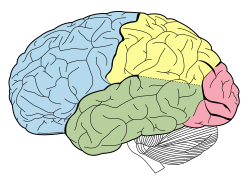
The frontal lobe is the largest lobe of the brain and makes up about a third of the surface area of each hemisphere. [3] On the lateral surface of each hemisphere, the central sulcus separates the frontal lobe from the parietal lobe. The lateral sulcus separates the frontal lobe from the temporal lobe.

The frontal lobe can be divided into a lateral, polar, orbital (above the orbit; also called basal or ventral), and medial part. Each of these parts consists of a particular gyrus:

The gyri are separated by sulci. E.g., the precentral gyrus is in front of the central sulcus, and behind the precentral sulcus. The superior and middle frontal gyri are divided by the superior frontal sulcus. The middle and inferior frontal gyri are divided by the inferior frontal sulcus.

In humans the frontal lobe reaches full maturity only after the 20s—the prefrontal cortex, in particular, continues in maturing till the second and third decades of life [5] —which, thereafter, marks the cognitive maturity associated with adulthood. A small amount of atrophy, however, is normal in the aging person's frontal lobe. Fjell, in 2009, studied atrophy of the brain in people aged 60–91 years. The 142 healthy participants were scanned using MRI. Their results were compared to those of 122 participants with Alzheimer's disease. A follow-up one year later showed there to have been a marked volumetric decline in those with Alzheimer's and a much smaller decline (averaging 0.5%) in the healthy group. [6] These findings corroborate those of Coffey, who in 1992 indicated that the frontal lobe decreases in volume approximately 0.5–1% per year. [7]

Function

The entirety of the frontal cortex can be considered the "action cortex", much as the posterior cortex is considered the "sensory cortex". It is devoted to action of one kind or another: skeletal movement, ocular movement, speech control, and the expression of emotions. In humans, the largest part of the frontal cortex, the prefrontal cortex (PFC), is responsible for internal, purposeful mental action, commonly called reasoning or prefrontal synthesis.

The function of the PFC involves the ability to project future consequences that result from current actions. PFC functions also include override and suppression of socially unacceptable responses as well as differentiation of tasks.

The PFC also plays an important part in integrating longer non-task based memories stored across the brain. These are often memories associated with emotions derived from input from the brain's limbic system. The frontal lobe modifies those emotions, generally to fit socially acceptable norms.

Psychological tests that measure frontal lobe function include finger tapping (as the frontal lobe controls voluntary movement), the Wisconsin Card Sorting Test, and measures of language, numeracy skills [8] and decision making [9] which are all controlled by frontal lobe.

Clinical significance

Damage

Damage to the frontal lobe can occur in a number of ways and result in many different consequences. Transient ischemic attacks (TIAs) also known as mini-strokes, and strokes are common causes of frontal lobe damage in older adults (65 and over). These strokes and mini-strokes can occur due to the blockage of blood flow to the brain or as a result of the rupturing of an aneurysm in a cerebral artery. Other ways in which injury can occur include traumatic brain injuries incurred following accidents, diagnoses such as Alzheimer's disease or Parkinson's disease (which cause dementia symptoms), and frontal lobe epilepsy (which can occur at any age). [10] Very often, frontal lobe damage is recognized in those with prenatal alcohol exposure.

Symptoms

Common effects of damage to the frontal lobe are varied. Patients who have experienced frontal lobe trauma may know the appropriate response to a situation but display inappropriate responses to those same situations in "real life". Similarly, emotions that are felt may not be expressed in the face or voice. For example, someone who is feeling happy would not smile, and the voice would be devoid of emotion. Along the same lines, though, the person may also exhibit excessive, unwarranted displays of emotion. Depression is common in stroke patients. Also common is a loss of or decrease in motivation. Someone might not want to carry out normal daily activities and would not feel "up to it". [10] Those who are close to the person who has experienced the damage may notice changes in behavior. [11] This personality change is characteristic of damage to the frontal lobe and was exemplified in the case of Phineas Gage. The frontal lobe is the same part of the brain that is responsible for executive functions such as planning for the future, judgment, decision-making skills, attention span, and inhibition. These functions can decrease drastically in someone whose frontal lobe is damaged. [10]

Consequences that are seen less frequently are also varied. Confabulation may be the most frequently indicated "less common" effect. In the case of confabulation, someone gives false information while maintaining the belief that it is the truth. In a small number of patients, uncharacteristic cheerfulness can be noted. This effect is seen mostly in patients with lesions to the right frontal portion of the brain. [10] [12]

Another infrequent effect is that of reduplicative paramnesia, in which patients believe that the location in which they currently reside is a replica of one located somewhere else. Similarly, those who experience Capgras syndrome after frontal lobe damage believe that an identical "replacement" has taken the identity of a close friend, relative, or other person and is posing as that person. This last effect is seen mostly in schizophrenic patients who also have a neurological disorder in the frontal lobe. [10] [13]

DNA damage

In the human frontal cortex, a set of genes undergo reduced expression after age 40 and especially after age 70. [14] This set includes genes that have key functions in synaptic plasticity important in learning and memory, vesicular transport and mitochondrial function. During aging, DNA damage is markedly increased in the promoters of the genes displaying reduced expression in the frontal cortex. In cultured human neurons, these promoters are selectively damaged by oxidative stress. [14]

Individuals with HIV associated neurocognitive disorders accumulate nuclear and mitochondrial DNA damage in the frontal cortex. [15]

Genetic

A report from the National Institute of Mental Health says a gene variant of (COMT) that reduces dopamine activity in the prefrontal cortex is related to poorer performance and inefficient functioning of that brain region during working memory, tasks, and to a slightly increased risk for schizophrenia. [16]

History

Psychosurgery

In the early 20th century, a medical treatment for mental illness, first developed by Portuguese neurologist Egas Moniz, involved damaging the pathways connecting the frontal lobe to the limbic system. A frontal lobotomy (sometimes called frontal leucotomy) successfully reduced distress but at the cost of often blunting the subject's emotions, volition and personality. The indiscriminate use of this psychosurgical procedure, combined with its severe side effects and a mortality rate of 7.4 to 17 per cent, [17] earned it a bad reputation. The frontal lobotomy has largely died out as a psychiatric treatment. More precise psychosurgical procedures are still used, although rarely. They may include anterior capsulotomy (bilateral thermal lesions of the anterior limbs of the internal capsule) or the bilateral cingulotomy (involving lesions of the anterior cingulate gyri) and might be used to treat otherwise untreatable obsessional disorders or clinical depression.

Theories of function

Theories of frontal lobe function can be separated into four categories:

Other theories include:

It may be highlighted that the theories described above differ in their focus on certain processes/systems or construct-lets. Stuss (1999) remarks that the question of homogeneity (single construct) or heterogeneity (multiple processes/systems) of function "may represent a problem of semantics and/or incomplete functional analysis rather than an unresolvable dichotomy" (p. 348). However, further research will show if a unified theory of frontal lobe function that fully accounts for the diversity of functions will be available.

Other primates

Many scientists had thought that the frontal lobe was disproportionately enlarged in humans compared to other primates. This was thought to be an important feature of human evolution and seen as the primary reason why human cognition differs from that of other primates. However, this view in relation to great apes has since been challenged by neuroimaging studies. Using magnetic resonance imaging to determine the volume of the frontal cortex in humans, all extant ape species and several monkey species, it was found that the human frontal cortex was not relatively larger than the cortex of other great apes but was relatively larger than the frontal cortex of lesser apes and the monkeys. [22] The higher cognition of the humans is instead seen to relate to a greater connectedness given by neural tracts that do not affect the cortical volume. [22] This is also evident in the pathways of the language network connecting the frontal and temporal lobes. [23]

See also

Related Research Articles

<span class="mw-page-title-main">Cingulate cortex</span> Part of the brain within the cerebral cortex

The cingulate cortex is a part of the brain situated in the medial aspect of the cerebral cortex. The cingulate cortex includes the entire cingulate gyrus, which lies immediately above the corpus callosum, and the continuation of this in the cingulate sulcus. The cingulate cortex is usually considered part of the limbic lobe.

<span class="mw-page-title-main">Brodmann area</span> Region of the brain

A Brodmann area is a region of the cerebral cortex, in the human or other primate brain, defined by its cytoarchitecture, or histological structure and organization of cells. The concept was first introduced by the German anatomist Korbinian Brodmann in the early 20th century. Brodmann mapped the human brain based on the varied cellular structure across the cortex and identified 52 distinct regions, which he numbered 1 to 52. These regions, or Brodmann areas, correspond with diverse functions including sensation, motor control, and cognition.

<span class="mw-page-title-main">Brodmann area 6</span>

Brodmann area 6 (BA6) is part of the frontal cortex in the human brain. Situated just anterior to the primary motor cortex (BA4), it is composed of the premotor cortex and, medially, the supplementary motor area (SMA). This large area of the frontal cortex is believed to play a role in planning complex, coordinated movements.

<span class="mw-page-title-main">Brodmann area 10</span> Brain area

Brodmann area 10 is the anterior-most portion of the prefrontal cortex in the human brain. BA10 was originally defined broadly in terms of its cytoarchitectonic traits as they were observed in the brains of cadavers, but because modern functional imaging cannot precisely identify these boundaries, the terms anterior prefrontal cortex, rostral prefrontal cortex and frontopolar prefrontal cortex are used to refer to the area in the most anterior part of the frontal cortex that approximately covers BA10—simply to emphasize the fact that BA10 does not include all parts of the prefrontal cortex.

<span class="mw-page-title-main">Brodmann area 11</span> Brain area

Brodmann area 11 is one of Brodmann's cytologically defined regions of the brain. It is in the orbitofrontal cortex which is above the eye sockets (orbitae). It is involved in decision making, processing rewards, and encoding new information into long-term memory.

<span class="mw-page-title-main">Inferior frontal gyrus</span> Part of the brains prefrontal cortex

The inferior frontal gyrus (IFG),, is the lowest positioned gyrus of the frontal gyri, of the frontal lobe, and is part of the prefrontal cortex.

<span class="mw-page-title-main">Brodmann area 4</span>

Brodmann area 4 refers to the primary motor cortex of the human brain. It is located in the posterior portion of the frontal lobe.

<span class="mw-page-title-main">Superior temporal gyrus</span> One of three gyri of the temporal lobe of the brain

The superior temporal gyrus (STG) is one of three gyri in the temporal lobe of the human brain, which is located laterally to the head, situated somewhat above the external ear.

<span class="mw-page-title-main">Precentral gyrus</span> Motor gyrus of the posterior frontal lobe of the brain

The precentral gyrus is a prominent gyrus on the surface of the posterior frontal lobe of the brain. It is the site of the primary motor cortex that in humans is cytoarchitecturally defined as Brodmann area 4.

<span class="mw-page-title-main">Middle cerebral artery</span> Paired artery that supplies blood to the cerebrum

The middle cerebral artery (MCA) is one of the three major paired cerebral arteries that supply blood to the cerebrum. The MCA arises from the internal carotid artery and continues into the lateral sulcus where it then branches and projects to many parts of the lateral cerebral cortex. It also supplies blood to the anterior temporal lobes and the insular cortices.

<span class="mw-page-title-main">Lobes of the brain</span> Parts of the cerebrum

The lobes of the brain are the major identifiable zones of the human cerebral cortex, and they comprise the surface of each hemisphere of the cerebrum. The two hemispheres are roughly symmetrical in structure, and are connected by the corpus callosum. They traditionally have been divided into four lobes, but are today considered as having six lobes each. The lobes are large areas that are anatomically distinguishable, and are also functionally distinct to some degree. Each lobe of the brain has numerous ridges, or gyri, and furrows, the sulci that constitute further subzones of the cortex. The expression "lobes of the brain" usually refers only to those of the cerebrum, not to the distinct areas of the cerebellum.

<span class="mw-page-title-main">Frontal gyri</span> Four gyri of the frontal lobe in the brain

The frontal gyri are four gyri of the frontal lobe in the brain. These are four horizontally oriented, parallel convolutions, of the frontal lobe. The other main gyrus of the frontal lobe is the precentral gyrus which is vertically oriented, and runs parallel with the precentral sulcus.

<span class="mw-page-title-main">Orbitofrontal cortex</span> Region of the prefrontal cortex of the brain

The orbitofrontal cortex (OFC) is a prefrontal cortex region in the frontal lobes of the brain which is involved in the cognitive process of decision-making. In non-human primates it consists of the association cortex areas Brodmann area 11, 12 and 13; in humans it consists of Brodmann area 10, 11 and 47.

<span class="mw-page-title-main">Frontal lobe disorder</span> Brain disorder

Frontal lobe disorder, also frontal lobe syndrome, is an impairment of the frontal lobe of the brain due to disease or frontal lobe injury. The frontal lobe plays a key role in executive functions such as motivation, planning, social behaviour, and speech production. Frontal lobe syndrome can be caused by a range of conditions including head trauma, tumours, neurodegenerative diseases, neurodevelopmental disorders, neurosurgery and cerebrovascular disease. Frontal lobe impairment can be detected by recognition of typical signs and symptoms, use of simple screening tests, and specialist neurological testing.

<span class="mw-page-title-main">Inferior temporal gyrus</span> One of three gyri of the temporal lobe of the brain

The inferior temporal gyrus is one of three gyri of the temporal lobe and is located below the middle temporal gyrus, connected behind with the inferior occipital gyrus; it also extends around the infero-lateral border on to the inferior surface of the temporal lobe, where it is limited by the inferior sulcus. This region is one of the higher levels of the ventral stream of visual processing, associated with the representation of objects, places, faces, and colors. It may also be involved in face perception, and in the recognition of numbers and words.

<span class="mw-page-title-main">Sulcus (neuroanatomy)</span> Fold in the surface of the brain

In neuroanatomy, a sulcus is a depression or groove in the cerebral cortex. It surrounds a gyrus, creating the characteristic folded appearance of the brain in humans and other mammals. The larger sulci are usually called fissures.

<span class="mw-page-title-main">Superior longitudinal fasciculus</span> Association fiber tract of the brain

The superior longitudinal fasciculus (SLF) is an association tract in the brain that is composed of three separate components. It is present in both hemispheres and can be found lateral to the centrum semiovale and connects the frontal, occipital, parietal, and temporal lobes. This bundle of tracts (fasciculus) passes from the frontal lobe through the operculum to the posterior end of the lateral sulcus where they either radiate to and synapse on neurons in the occipital lobe, or turn downward and forward around the putamen and then radiate to and synapse on neurons in anterior portions of the temporal lobe.

<span class="mw-page-title-main">Orbital part of inferior frontal gyrus</span>

The orbital part of inferior frontal gyrus also known as the pars orbitalis is the orbital part of the inferior frontal gyrus.

Emotional lateralization is the asymmetrical representation of emotional control and processing in the brain. There is evidence for the lateralization of other brain functions as well.

References

  1. Muzio, Bruno Di. "Frontal pole | Radiology Reference Article | Radiopaedia.org". radiopaedia.org.
  2. João, Rafael Batista; Filgueiras, Raquel Mattos (2018-10-03), Starcevic, Ana; Filipovic, Branislav (eds.), "Frontal Lobe: Functional Neuroanatomy of Its Circuitry and Related Disconnection Syndromes", Prefrontal Cortex, InTech, doi: 10.5772/intechopen.79571 , ISBN   978-1-78923-903-4, S2CID   149544570 , retrieved 2023-06-24
  3. 1 2 Carpenter, Malcolm (1985). Core text of neuroanatomy (3rd ed.). Williams & Wilkins. pp. 22–23. ISBN   978-0683014556.
  4. "Incoming Sensory Information - an overview | ScienceDirect Topics". www.sciencedirect.com. Retrieved 2023-03-31.
  5. Kolk, Sharon M.; Rakic, Pasko (January 2022). "Development of prefrontal cortex". Neuropsychopharmacology. 47 (1): 41–57. doi:10.1038/s41386-021-01137-9. ISSN   1740-634X. PMC   8511863 . PMID   34645980.
  6. Fjell AM, Walhovd KB, Fennema-Notestine C, McEvoy LK, Hagler DJ, Holland D, Brewer JB, Dale AM (December 2009). "One-year brain atrophy evident in healthy aging". The Journal of Neuroscience. 29 (48): 15223–31. doi:10.1523/JNEUROSCI.3252-09.2009. PMC   2827793 . PMID   19955375.
  7. Coffey CE, Wilkinson WE, Parashos IA, Soady SA, Sullivan RJ, Patterson LJ, Figiel GS, Webb MC, Spritzer CE, Djang WT (March 1992). "Quantitative cerebral anatomy of the aging human brain: a cross-sectional study using magnetic resonance imaging". Neurology. 42 (3 Pt 1): 527–36. doi:10.1212/wnl.42.3.527. PMID   1549213. S2CID   20481757.
  8. Kimberg DY, Farah MJ (December 1993). "A unified account of cognitive impairments following frontal lobe damage: the role of working memory in complex, organized behavior". Journal of Experimental Psychology. General. 122 (4): 411–28. doi:10.1037/0096-3445.122.4.411. PMID   8263463.
  9. Yang X, Gao M, Shi J, Ye H, Chen S (2017). "Modulating the Activity of the DLPFC and OFC Has Distinct Effects on Risk and Ambiguity Decision-Making: A tDCS Study". Frontiers in Psychology. 8: 1417. doi: 10.3389/fpsyg.2017.01417 . PMC   5572270 . PMID   28878714.
  10. 1 2 3 4 5 Stuss DT, Gow CA, Hetherington CR (June 1992). ""No longer Gage": frontal lobe dysfunction and emotional changes". Journal of Consulting and Clinical Psychology. 60 (3): 349–59. doi:10.1037/0022-006X.60.3.349. PMID   1619089.
  11. Rowe AD, Bullock PR, Polkey CE, Morris RG (March 2001). ""Theory of mind" impairments and their relationship to executive functioning following frontal lobe excisions". Brain. 124 (Pt 3): 600–16. doi: 10.1093/brain/124.3.600 . PMID   11222459.
  12. Robinson RG, Kubos KL, Starr LB, Rao K, Price TR (March 1984). "Mood disorders in stroke patients. Importance of location of lesion". Brain. 107 ( Pt 1) (1): 81–93. doi:10.1093/brain/107.1.81. PMID   6697163.
  13. Durani SK, Ford R, Sajjad SH (September 1991). "Capgras syndrome associated with a frontal lobe tumour". Irish Journal of Psychological Medicine. 8 (2): 135–6. doi:10.1017/S0790966700015093. S2CID   74081936.
  14. 1 2 Lu T, Pan Y, Kao SY, Li C, Kohane I, Chan J, Yankner BA (June 2004). "Gene regulation and DNA damage in the ageing human brain". Nature. 429 (6994): 883–91. Bibcode:2004Natur.429..883L. doi:10.1038/nature02661. PMID   15190254. S2CID   1867993.
  15. Zhang Y, Wang M, Li H, Zhang H, Shi Y, Wei F, Liu D, Liu K, Chen D (June 2012). "Accumulation of nuclear and mitochondrial DNA damage in the frontal cortex cells of patients with HIV-associated neurocognitive disorders". Brain Research. 1458: 1–11. doi:10.1016/j.brainres.2012.04.001. PMID   22554480. S2CID   23744888.
  16. "Gene Slows Frontal Lobes, Boosts Schizophrenia Risk". National Institute of Mental Health. May 29, 2001. Archived from the original on 2015-04-04. Retrieved 2013-06-20.
  17. Ogren K, Sandlund M (2007). "Lobotomy at a state mental hospital in Sweden. A survey of patients operated on during the period 1947–1958". Nordic Journal of Psychiatry. 61 (5): 355–62. doi:10.1080/08039480701643498. PMID   17990197. S2CID   26307989.
  18. (Burgess, 2003, p. 309).
  19. (Burgess, 2003, p. 310).
  20. (Stuss, 1999, p. 348; cf. Burgess & Simons, 2005).
  21. (cf. Burgess & Simons, 2005).
  22. 1 2 Semendeferi K, Lu A, Schenker N, Damasio H (March 2002). "Humans and great apes share a large frontal cortex". Nature Neuroscience. 5 (3): 272–6. doi:10.1038/nn814. PMID   11850633. S2CID   5921065.
  23. Friederici AD (April 2009). "Pathways to language: fiber tracts in the human brain". Trends in Cognitive Sciences. 13 (4): 175–81. doi:10.1016/j.tics.2009.01.001. PMID   19223226. S2CID   15609709.

Further reading