| Cerebral hemisphere | |
|---|---|
 Human brain seen from front | |
 Right cerebral hemisphere Left cerebral hemisphere | |
| Details | |
| Identifiers | |
| Latin | hemisphaerium cerebri |
| NeuroNames | 241 |
| NeuroLex ID | birnlex_1796 |
| TA98 | A14.1.09.002 |
| TA2 | 5418 |
| FMA | 61817 |
| Anatomical terms of neuroanatomy | |
The cerebrum, or the largest part of the vertebrate brain, is made up of two cerebral hemispheres. The deep groove known as the longitudinal fissure divides the cerebrum into the left and right hemispheres, but the hemispheres remain united by the corpus callosum, a large bundle of nerve fibers in the middle of the brain whose primary function is to integrate sensory and motor signals between the hemispheres. In eutherian (placental) mammals, other bundles of nerve fibers like the corpus callosum exist, including the anterior commissure, the posterior commissure, and the fornix, but compared with the corpus callosum, they are much smaller in size.
Contents
- Structure
- Poles
- Composition
- Development
- Function
- Hemisphere lateralization
- Clinical significance
- Additional images
- References
Broadly, the hemispheres are made up of two types of tissues. The thin outer layer of the cerebral hemispheres is made up of gray matter, composed of neuronal cell bodies, dendrites, and synapses; this outer layer constitutes the cerebral cortex (cortex is Latin for "bark of a tree"). Below that is the larger inner layer of white matter, composed of axons and myelin.
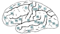
Each hemisphere is further subdivided into a frontal, parietal, occipital, and temporal lobe. The central sulcus is a prominent fissure that separates both the frontal lobe from the parietal lobe and the primary motor cortex from the primary somatosensory cortex. Three of the four lobes also have "poles": the occipital pole , the frontal pole , and the temporal pole .
The two cerebral hemispheres are nicely macroscopic mirror images of each other, with subtle anatomical differences between them, such as the Yakovlevian torque that is sometimes seen in the human brain. Nevertheless, on a microscopic level, the functions of cells, the quantities of neurotransmitters, and the types of receptors between the hemispheres is markedly asymmetrical. [1] [2] While some of these hemispheric distribution differences are consistent across human beings, or even across some species, many observable distribution differences vary from individual to individual within a given species.