Brodmann area 44

Last updated
Brodmann area 44
Ba44.png
Brodmann Cytoarchitectonics 44.png
Details
Artery middle cerebral artery
Identifiers
Latin area opercularis
NeuroLex ID birnlex_1776
FMA 68641
Anatomical terms of neuroanatomy

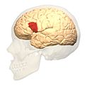
Brodmann area 44, or BA44, is part of the frontal cortex in the human brain. Situated just anterior to premotor cortex (BA6) and on the lateral surface, inferior to BA9.

Contents

This area is also known as pars opercularis (of the inferior frontal gyrus), and it refers to a subdivision of the cytoarchitecturally defined frontal region of cerebral cortex. In the human it corresponds approximately to the opercular part of the inferior frontal gyrus. Thus, it is bounded caudally by the inferior precentral sulcus (H) and rostrally by the anterior ascending limb of lateral sulcus (H). It surrounds the diagonal sulcus (H). In the depth of the lateral sulcus it borders on the insula. Cytoarchitectonically it is bounded caudally and dorsally by the agranular frontal area 6, dorsally by the granular frontal area 9 and rostrally by the triangular part of inferior frontal gyrus (Brodmann area 45 BA 45).

Functions

Together with left-hemisphere BA45, the left hemisphere [1] BA44 comprises Broca's area, a region involved in semantic tasks. Some data suggest that BA44 is more involved in the motor aspect of speech. Some recent findings also suggest the implication of this region in music perception. [2] Recent neuroimaging studies show BA44 involvement in selective response suppression in go/no-go tasks and is therefore believed to play an important role in the suppression of response tendencies. [3] This may apply mainly to the right BA44. [4] Neuroimaging studies also demonstrate that area 44 is related to hand movements. [5]

The presence of mirror neurons in Broca's area suggests language evolved from a gesture imitating system. Broca's area is also involved with theory of mind (ToM), which is the ability to understand the mental state of others through observation, inferring, and projecting. [6]

Trivia

Scott Flansburg of San Diego, California is a "human calculator" who can perform complex arithmetic in his head. Profiled on the TV show Stan Lee's Superhumans, his brain was scanned using fMRI while doing complex calculations, which showed brain activity in this region was absent. Instead there was activity somewhat higher than that of area 44 and closer to the motor cortex. [7]

Image

See also

Notes and references

  1. In 95.5% of right-handers and 61.4% of left-handers, therefore about 92% of the clinical population, speech is lateralised in the left hemisphere.
  2. Brown, Steven; Michael J. Martinez & Lawrence M. Parsons (2006). "Music and language side by side in the brain: a PET study of the generation of melodies and sentences". European Journal of Neuroscience. 23 (10): 2791–2803. CiteSeerX   10.1.1.530.5981 . doi:10.1111/j.1460-9568.2006.04785.x. PMID   16817882. S2CID   15189129.
  3. Forstmann, Birte U.; Van den Wildenberg, Wery P.M. and Ridderinkhof, Richard K (2008). Neural Mechanisms, Temporal Dynamics, and Individual Differences in Interference Control. Journal of Cognitive Neuroscience 20:10, pp. 1854-65
  4. Neef, NE; Bütfering, C; Anwander, A; Friederici, AD; Paulus, W; Sommer, M (2016). "Left posterior-dorsal area 44 couples with parietal areas to promote speech fluency, while right area 44 activity promotes the stopping of motor responses". NeuroImage. 142: 628–644. doi: 10.1016/j.neuroimage.2016.08.030 . hdl: 11858/00-001M-0000-002B-2E34-C . PMID   27542724. S2CID   14864376.
  5. Rizzolatti, G.; Fogassi, L.; Gallese, V. (2002). "Motor and cognitive functions of the ventral premotor cortex". Current Opinion in Neurobiology. 12 (2): 149–154. doi:10.1016/S0959-4388(02)00308-2. PMID   12015230. S2CID   17840974.
  6. Rajmohan, V (2007). "Mirror neuron system". Indian J Psychiatry. 49 (10): 66–69. doi: 10.4103/0019-5545.31522 . PMC   2900004 . PMID   20640069.
  7. "Electro Man: Episode 101". Stan Lee's Superhumans. August 5, 2011.

Related Research Articles

<span class="mw-page-title-main">Broca's area</span> Speech production region in the dominant hemisphere of the hominid brain

Broca's area, or the Broca area, is a region in the frontal lobe of the dominant hemisphere, usually the left, of the brain with functions linked to speech production.

<span class="mw-page-title-main">Cingulate cortex</span> Part of the brain within the cerebral cortex

The cingulate cortex is a part of the brain situated in the medial aspect of the cerebral cortex. The cingulate cortex includes the entire cingulate gyrus, which lies immediately above the corpus callosum, and the continuation of this in the cingulate sulcus. The cingulate cortex is usually considered part of the limbic lobe.

<span class="mw-page-title-main">Brodmann area</span> Region of the brain

A Brodmann area is a region of the cerebral cortex, in the human or other primate brain, defined by its cytoarchitecture, or histological structure and organization of cells. The concept was first introduced by the German anatomist Korbinian Brodmann in the early 20th century. Brodmann mapped the human brain based on the varied cellular structure across the cortex and identified 52 distinct regions, which he numbered 1 to 52. These regions, or Brodmann areas, correspond with diverse functions including sensation, motor control, and cognition.

<span class="mw-page-title-main">Parietal lobe</span> Part of the brain responsible for sensory input and some language processing

The parietal lobe is one of the four major lobes of the cerebral cortex in the brain of mammals. The parietal lobe is positioned above the temporal lobe and behind the frontal lobe and central sulcus.

<span class="mw-page-title-main">Precuneus</span> Region of the parietal lobe of the brain

In neuroanatomy, the precuneus is the portion of the superior parietal lobule on the medial surface of each brain hemisphere. It is located in front of the cuneus. The precuneus is bounded in front by the marginal branch of the cingulate sulcus, at the rear by the parieto-occipital sulcus, and underneath by the subparietal sulcus. It is involved with episodic memory, visuospatial processing, reflections upon self, and aspects of consciousness.

<span class="mw-page-title-main">Brodmann area 6</span>

Brodmann area 6 (BA6) is part of the frontal cortex in the human brain. Situated just anterior to the primary motor cortex (BA4), it is composed of the premotor cortex and, medially, the supplementary motor area (SMA). This large area of the frontal cortex is believed to play a role in planning complex, coordinated movements.

<span class="mw-page-title-main">Brodmann area 10</span> Brain area

Brodmann area 10 is the anterior-most portion of the prefrontal cortex in the human brain. BA10 was originally defined broadly in terms of its cytoarchitectonic traits as they were observed in the brains of cadavers, but because modern functional imaging cannot precisely identify these boundaries, the terms anterior prefrontal cortex, rostral prefrontal cortex and frontopolar prefrontal cortex are used to refer to the area in the most anterior part of the frontal cortex that approximately covers BA10—simply to emphasize the fact that BA10 does not include all parts of the prefrontal cortex.

<span class="mw-page-title-main">Brodmann area 45</span> Brain area

Brodmann area 45 (BA45), is part of the frontal cortex in the human brain. It is situated on the lateral surface, inferior to BA9 and adjacent to BA46.

<span class="mw-page-title-main">Brodmann area 47</span> Brain area

Brodmann area 47, or BA47, is part of the frontal cortex in the human brain. It curves from the lateral surface of the frontal lobe into the ventral (orbital) frontal cortex. It is below areas BA10 and BA45, and beside BA11. This cytoarchitectonic region most closely corresponds to the gyral region the orbital part of inferior frontal gyrus, although these regions are not equivalent. Pars orbitalis is not based on cytoarchitectonic distinctions, and rather is defined according to gross anatomical landmarks. Despite a clear distinction, these two terms are often used liberally in peer-reviewed research journals.

<span class="mw-page-title-main">Brodmann area 40</span> Part of the parietal cortex in the human brain

Brodmann area 40 (BA40) is part of the parietal cortex in the human brain. The inferior part of BA40 is in the area of the supramarginal gyrus, which lies at the posterior end of the lateral fissure, in the inferior lateral part of the parietal lobe.

<span class="mw-page-title-main">Brodmann area 11</span> Brain area

Brodmann area 11 is one of Brodmann's cytologically defined regions of the brain. It is in the orbitofrontal cortex which is above the eye sockets (orbitae). It is involved in decision making, processing rewards, and encoding new information into long-term memory.

<span class="mw-page-title-main">Inferior frontal gyrus</span> Part of the brains prefrontal cortex

The inferior frontal gyrus (IFG),, is the lowest positioned gyrus of the frontal gyri, of the frontal lobe, and is part of the prefrontal cortex.

<span class="mw-page-title-main">Arcuate fasciculus</span> Neural pathway connecting Brocas area and Wernickes area

In neuroanatomy, the arcuate fasciculus is a bundle of axons that generally connects the Broca's area and the Wernicke's area in the brain. It is an association fiber tract connecting caudal temporal cortex and inferior frontal lobe.

<span class="mw-page-title-main">Lobes of the brain</span> Parts of the cerebrum

The lobes of the brain are the major identifiable zones of the human cerebral cortex, and they comprise the surface of each hemisphere of the cerebrum. The two hemispheres are roughly symmetrical in structure, and are connected by the corpus callosum. They traditionally have been divided into four lobes, but are today considered as having six lobes each. The lobes are large areas that are anatomically distinguishable, and are also functionally distinct to some degree. Each lobe of the brain has numerous ridges, or gyri, and furrows, the sulci that constitute further subzones of the cortex. The expression "lobes of the brain" usually refers only to those of the cerebrum, not to the distinct areas of the cerebellum.

<span class="mw-page-title-main">Premotor cortex</span>

The premotor cortex is an area of the motor cortex lying within the frontal lobe of the brain just anterior to the primary motor cortex. It occupies part of Brodmann's area 6. It has been studied mainly in primates, including monkeys and humans. The functions of the premotor cortex are diverse and not fully understood. It projects directly to the spinal cord and therefore may play a role in the direct control of behavior, with a relative emphasis on the trunk muscles of the body. It may also play a role in planning movement, in the spatial guidance of movement, in the sensory guidance of movement, in understanding the actions of others, and in using abstract rules to perform specific tasks. Different subregions of the premotor cortex have different properties and presumably emphasize different functions. Nerve signals generated in the premotor cortex cause much more complex patterns of movement than the discrete patterns generated in the primary motor cortex.

<span class="mw-page-title-main">Superior longitudinal fasciculus</span> Association fiber tract of the brain

The superior longitudinal fasciculus (SLF) is an association tract in the brain that is composed of three separate components. It is present in both hemispheres and can be found lateral to the centrum semiovale and connects the frontal, occipital, parietal, and temporal lobes. This bundle of tracts (fasciculus) passes from the frontal lobe through the operculum to the posterior end of the lateral sulcus where they either radiate to and synapse on neurons in the occipital lobe, or turn downward and forward around the putamen and then radiate to and synapse on neurons in anterior portions of the temporal lobe.

<span class="mw-page-title-main">Orbital part of inferior frontal gyrus</span>

The orbital part of inferior frontal gyrus also known as the pars orbitalis is the orbital part of the inferior frontal gyrus.

Nonprimary motor cortex is a functionally defined portion of the frontal lobe. It includes two subdivisions, the premotor cortex and the supplementary motor cortex. Largely coincident with the cytoarchitecturally defined area 6 of Brodmann (human), it is located primarily in the rostral portion of the precentral gyrus and caudal portions of the superior frontal gyrus and the middle frontal gyrus, It aids in cerebral control of movement. Anatomically speaking, several nonmprimary areas exist, and make direct connections with the spinal cord.

Social cognitive neuroscience is the scientific study of the biological processes underpinning social cognition. Specifically, it uses the tools of neuroscience to study "the mental mechanisms that create, frame, regulate, and respond to our experience of the social world". Social cognitive neuroscience uses the epistemological foundations of cognitive neuroscience, and is closely related to social neuroscience. Social cognitive neuroscience employs human neuroimaging, typically using functional magnetic resonance imaging (fMRI). Human brain stimulation techniques such as transcranial magnetic stimulation and transcranial direct-current stimulation are also used. In nonhuman animals, direct electrophysiological recordings and electrical stimulation of single cells and neuronal populations are utilized for investigating lower-level social cognitive processes.