Occipital gyri

Last updated
Occipital gyri
OccCapts.png
Gyri of the occipital lobe shown on the right diagram
Details
System Visual System
LocationCerebrum
Identifiers
Latin gyri occipitalis
Anatomical terms of neuroanatomy

The occipital gyri (OcG) are three gyri in parallel, along the lateral portion of the occipital lobe, also referred to as a composite structure in the brain. [1] [2] The gyri are the superior occipital gyrus, the middle occipital gyrus, and the inferior occipital gyrus, and these are also known as the occipital face area. [1] The superior and inferior occipital sulci separates the three occipital gyri. [3]

Contents

The intraoccipital sulcus, also known as the superior occipital sulcus, stems from the intraparietal sulcus and continues until the sulcus reaches the transverse occipital sulcus, separating the superior occipital gyrus from the middle occipital gyrus. The transverse occipital sulcus comes down along the lateral occipital surface or the inferior occipital sulcus. [4]

Structural anatomy

Diagram of gyri of brain viewed on lateral hemisphere. Occipital gyri shown lower right. Gyri of lateral cortex.png
Diagram of gyri of brain viewed on lateral hemisphere. Occipital gyri shown lower right.

The border between the occipital lobe and the parietal and temporal lobes is characterized by different gyri: the superior occipital gyrus (also known as gyrus occipitalis superior), middle occipital gyrus (or gyrus occipitalis medius), inferior occipital gyrus (or gyrus occipitalis inferior), and descending occipital gyrus (gyrus occipitalis descendens). [5]

Function

The occipital complex is primarily responsible for object recognition, including the functional properties and our perception of said objects. [6] The middle occipital gyrus (MOG) was observed in a study of the early blind, which showed that it was activated more during spatial than nonspatial tactile and auditory tasks. [7] Early blind people have an occipital cortex that is incorporates more senses than people with standard vision, but the MOG still keeps its functional role in processing space around a person. [7]

The lingual gyrus (also known as medial occipitotemporal gyrus) has been studied and found to be included in processing overall shapes, rather than the individual components that make up a shape. [8] This shows that the lingual gyrus is active during visual processing. [8]

The inferior occipital gyrus has been found to be related to the visual function of processing faces. The IOG is connected to the amygdala via white matter connectivity. [9] This allows the IOG to form a network for facial recognition with the amygdala. [9]

Development

The occipital lobe becomes distinct at 18 weeks gestation, but the gyri are not clear until many weeks later. [10] During development, the occipital lobe develops a lingual gyrus at 27 weeks of gestation. [10] Secondary gyri develop by 30 weeks, and tertiary gyri develop during 40 to 42 weeks of gestation. [10] The superior and inferior occipital gyri develop at the same time, usually shown somewhere between week 24 and 27 in brain development. [10] Due to the unclear distinction in early neuroscience research as to whether there are two or three occipital gyri, there is not any data on when the middle occipital gyrus starts its formation, but it is likely at the same time.

Related Research Articles

Parietal lobe Part of the brain responsible for sensory input and some language processing

The parietal lobe is one of the four major lobes of the cerebral cortex in the brain of mammals. The parietal lobe is positioned above the temporal lobe and behind the frontal lobe and central sulcus.

Frontal lobe Part of the brain

The frontal lobe is the largest of the four major lobes of the brain in mammals, and is located at the front of each cerebral hemisphere. It is parted from the parietal lobe by a groove between tissues called the central sulcus and from the temporal lobe by a deeper groove called the lateral sulcus. The most anterior rounded part of the frontal lobe is known as the frontal pole, one of the three poles of the cerebrum.

Occipital lobe Part of the brain at the back of the head

The occipital lobe is one of the four major lobes of the cerebral cortex in the brain of mammals. The name derives from its position at the back of the head, from the Latin ob, "behind," and caput, "the head."

Fusiform gyrus Gyrus of the temporal and occipital lobes of the brain

The fusiform gyrus, also known as the lateral occipitotemporal gyrus,is part of the temporal lobe and occipital lobe in Brodmann area 37. The fusiform gyrus is located between the lingual gyrus and parahippocampal gyrus above, and the inferior temporal gyrus below. Though the functionality of the fusiform gyrus is not fully understood, it has been linked with various neural pathways related to recognition. Additionally, it has been linked to various neurological phenomena such as synesthesia, dyslexia, and prosopagnosia.

Lateral sulcus

The lateral sulcus is one of the most prominent features of the human brain. The lateral sulcus is a deep fissure in each hemisphere that separates the frontal and parietal lobes from the temporal lobe. The insular cortex lies deep within the lateral sulcus.

Inferior frontal gyrus Part of the brains prefrontal cortex

The inferior frontal gyrus(IFG), (gyrus frontalis inferior), is the lowest positioned gyrus of the frontal gyri, of the frontal lobe, and is part of the prefrontal cortex.

Superior temporal gyrus One of three gyri of the temporal lobe of the brain

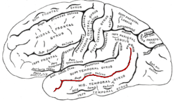
The superior temporal gyrus (STG) is one of three gyri in the temporal lobe of the human brain, which is located laterally to the head, situated somewhat above the external ear.

Angular gyrus Gyrus of the parietal lobe of the brain

The angular gyrus is a region of the brain lying mainly in the posteroinferior region of the parietal lobe, occupying the posterior part of the inferior parietal lobule. It represents the Brodmann area 39.

Gyrus

In neuroanatomy, a gyrus is a ridge on the cerebral cortex. It is generally surrounded by one or more sulci. Gyri and sulci create the folded appearance of the brain in humans and other mammals.

Lobes of the brain Parts of the cerebrum

The lobes of the brain are the major identifiable zones of the cerebral cortex, and they comprise the surface of each hemisphere of the cerebrum. The two hemispheres, which are only roughly symmetrical in structure, are today considered as having six lobes each. The lobes are large areas that are anatomically distinguishable, and are also functionally distinct to some degree. Each lobe of the brain has numerous ridges, or gyri, and furrows, the sulci that constitute further subzones of the cortex. The expression "lobes of the brain" usually refers only to those of the cerebrum, not to the distinct areas of the cerebellum.

Frontal gyri Four gyri of the frontal lobe in the brain

The frontal gyri are four gyri of the frontal lobe in the brain. These are four horizontally oriented, parallel convolutions, of the frontal lobe. The other main gyrus of the frontal lobe is the precentral gyrus which is vertically oriented, and runs parallel with the precentral sulcus.

Middle temporal gyrus One of three gyri of the temporal lobe of the brain

Middle temporal gyrus is a gyrus in the brain on the temporal lobe. It is located between the superior temporal gyrus and inferior temporal gyrus. It corresponds largely to Brodmann area 21.

Inferior temporal gyrus One of three gyri of the temporal lobe of the brain

The inferior temporal gyrus is one of three gyri of the temporal lobe and is located below the middle temporal gyrus, connected behind with the inferior occipital gyrus; it also extends around the infero-lateral border on to the inferior surface of the temporal lobe, where it is limited by the inferior sulcus. This region is one of the higher levels of the ventral stream of visual processing, associated with the representation of objects, places, faces, and colors. It may also be involved in face perception, and in the recognition of numbers.

Superior parietal lobule

The superior parietal lobule is bounded in front by the upper part of the postcentral sulcus, but is usually connected with the postcentral gyrus above the end of the sulcus. The superior parietal lobule contains Brodmann's areas 5 and 7.

Inferior parietal lobule Portion of the parietal lobe of the brain

The inferior parietal lobule lies below the horizontal portion of the intraparietal sulcus, and behind the lower part of the postcentral sulcus. Also known as Geschwind's territory after Norman Geschwind, an American neurologist, who in the early 1960s recognised its importance. It is a part of the parietal lobe.

Lingual gyrus Gyrus of the occipital lobe of the brain

The lingual gyrus, also known as the medialoccipitotemporal gyrus, is a brain structure that is linked to processing vision, especially related to letters. It is thought to also play a role in analysis of logical conditions and encoding visual memories. It is named after its shape, which is somewhat similar to a tongue. Contrary to the name, the region has little to do with speech.

Sulcus (neuroanatomy) Fold in the surface of the brain

In neuroanatomy, a sulcus is a depression or groove in the cerebral cortex. It surrounds a gyrus, creating the characteristic folded appearance of the brain in humans and other mammals. The larger sulci are usually called fissures.

Superior temporal sulcus Part of the brains temporal lobe

The superior temporal sulcus (STS) is the sulcus separating the superior temporal gyrus from the middle temporal gyrus in the temporal lobe of the brain. A sulcus is a deep groove that curves into the largest part of the brain, the cerebrum, and a gyrus is a ridge that curves outward of the cerebrum.

Neuroscience of sex differences Characteristics of the brain that differentiate the male brain and the female brain

The neuroscience of sex differences is the study of characteristics that separate the male and female brain. Psychological sex differences are thought by some to reflect the interaction of genes, hormones, and social learning on brain development throughout the lifespan.

References

  1. 1 2 Albohn DN, Adams Jr RB (January 2016). "Social Vision: At the Intersection of Vision and Person Perception". Neuroimaging Personality, Social Cognition, and Character. pp. 159–186. doi:10.1016/B978-0-12-800935-2.00008-7. ISBN   978-0-12-800935-2.
  2. "Occipital gyri". BrainInfo. University of Washington.
  3. Duvernoy H (May 2007). "Chapter 3 - Brain Anatomy". Magnetic Resonance in Epilepsy (2nd ed.). pp. 29–97. doi:10.1016/B978-012431152-7/50007-0. ISBN   978-0-12-431152-7.
  4. Alves RV, Ribas GC, Párraga RG, de Oliveira E (May 2012). "The occipital lobe convexity sulci and gyri". Journal of Neurosurgery. 116 (5): 1014–23. doi:10.3171/2012.1.JNS11978. PMID   22339163.
  5. Ten Donkelaar HJ, Tzourio-Mazoyer N, Mai JK (2018-11-19). "Toward a Common Terminology for the Gyri and Sulci of the Human Cerebral Cortex". Frontiers in Neuroanatomy. 12: 93. doi: 10.3389/fnana.2018.00093 . PMC   6252390 . PMID   30510504.
  6. Grill-Spector K, Kourtzi Z, Kanwisher N (2001). "The lateral occipital complex and its role in object recognition". Vision Research. 41 (10–11): 1409–22. doi:10.1016/s0042-6989(01)00073-6. PMID   11322983. S2CID   8100507.
  7. 1 2 Renier LA, Anurova I, De Volder AG, Carlson S, VanMeter J, Rauschecker JP (October 2010). "Preserved functional specialization for spatial processing in the middle occipital gyrus of the early blind". Neuron. 68 (1): 138–48. doi:10.1016/j.neuron.2010.09.021. PMC   2951740 . PMID   20920797.
  8. 1 2 Mechelli A, Humphreys GW, Mayall K, Olson A, Price CJ (September 2000). "Differential effects of word length and visual contrast in the fusiform and lingual gyri during reading". Proceedings. Biological Sciences. 267 (1455): 1909–13. doi:10.1098/rspb.2000.1229. PMC   1690747 . PMID   11052544.
  9. 1 2 Sato W, Kochiyama T, Uono S, Matsuda K, Usui K, Usui N, et al. (September 2017). "Bidirectional electric communication between the inferior occipital gyrus and the amygdala during face processing". Human Brain Mapping. 38 (9): 4511–4524. doi: 10.1002/hbm.23678 . PMC   6867177 . PMID   28573679.
  10. 1 2 3 4 Chi JG, Dooling EC, Gilles FH (January 1977). "Gyral development of the human brain". Annals of Neurology. 1 (1): 86–93. doi:10.1002/ana.410010109. PMID   560818. S2CID   22434544.