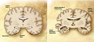
The hippocampus is a major component of the brain of humans and other vertebrates. Humans and other mammals have two hippocampi, one in each side of the brain. The hippocampus is part of the limbic system, and plays important roles in the consolidation of information from short-term memory to long-term memory, and in spatial memory that enables navigation. The hippocampus is located under the cerebral cortex in the allocortex, and in primates it is in the medial temporal lobe. It contains two main interlocking parts: the hippocampus proper and the dentate gyrus.

The cingulate cortex is a part of the brain situated in the medial aspect of the cerebral cortex. The cingulate cortex includes the entire cingulate gyrus, which lies immediately above the corpus callosum, and the continuation of this in the cingulate sulcus. The cingulate cortex is usually considered part of the limbic lobe.

The temporal lobe is involved in processing sensory input into derived meanings for the appropriate retention of visual memory, language comprehension, and emotion association.

The frontal lobe is the largest of the four major lobes of the brain in mammals, and is located at the front of each hemisphere. It is separated from the parietal lobe by a groove between tissues called the central sulcus, and from the temporal lobe by a deeper groove called the lateral sulcus. The most anterior rounded part of the frontal lobe is known as the frontal pole, one of the three poles of the cerebrum.

The piriform cortex, or pyriform cortex, is a region in the brain, part of the rhinencephalon situated in the cerebrum. The function of the piriform cortex relates to the sense of smell.

The olfactory system, or sense of smell, is the part of the sensory system used for smelling (olfaction). Most mammals and reptiles have a main olfactory system and an accessory olfactory system. The main olfactory system detects airborne substances, while the accessory system senses fluid-phase stimuli.

The fusiform gyrus, also known as the lateral occipitotemporal gyrus, is part of the temporal lobe and occipital lobe in Brodmann area 37. The fusiform gyrus is located between the lingual gyrus and parahippocampal gyrus above, and the inferior temporal gyrus below. Though the functionality of the fusiform gyrus is not fully understood, it has been linked with various neural pathways related to recognition. Additionally, it has been linked to various neurological phenomena such as synesthesia, dyslexia, and prosopagnosia.
The limbic lobe is an arc-shaped region of cortex on the medial surface of each cerebral hemisphere of the mammalian brain, consisting of parts of the frontal, parietal and temporal lobes. The term is ambiguous, with some authors including the paraterminal gyrus, the subcallosal area, the cingulate gyrus, the parahippocampal gyrus, the dentate gyrus, the hippocampus and the subiculum; while the Terminologia Anatomica includes the cingulate sulcus, the cingulate gyrus, the isthmus of cingulate gyrus, the fasciolar gyrus, the parahippocampal gyrus, the parahippocampal sulcus, the dentate gyrus, the fimbrodentate sulcus, the fimbria of hippocampus, the collateral sulcus, and the rhinal sulcus, and omits the hippocampus.

In neuroanatomy, a gyrus is a ridge on the cerebral cortex. It is generally surrounded by one or more sulci. Gyri and sulci create the folded appearance of the brain in humans and other mammals.

In neuroanatomy, the cingulum is a collection of white matter, fibers projecting from the cingulate gyrus to the entorhinal cortex in the brain, allowing for communication between components of the limbic system. It forms the white matter core of the cingulate gyrus, following it from the subcallosal gyrus of the frontal lobe beneath the rostrum of corpus callosum to the parahippocampal gyrus and uncus of the temporal lobe.

The subiculum is the most inferior component of the hippocampal formation. It lies between the entorhinal cortex and the CA1 subfield of the hippocampus proper.
The lobes of the brain were originally a purely anatomical classification, but have been shown also to be related to different brain functions. The cerebrum, the largest portion of the human brain, is divided into lobes, but so is the cerebellum. If not specified, the expression "lobes of the brain" refers to the cerebrum.

The inferior temporal gyrus is placed below the middle temporal gyrus, and is connected behind with the inferior occipital gyrus; it also extends around the infero-lateral border on to the inferior surface of the temporal lobe, where it is limited by the inferior sulcus. This region is one of the higher levels of the ventral stream of visual processing, associated with the representation of complex object features, such as global shape. It may also be involved in face perception, and in the recognition of numbers.

The posterior cingulate cortex (PCC) is the caudal part of the cingulate cortex, located posterior to the anterior cingulate cortex. This is the upper part of the "limbic lobe". The cingulate cortex is made up of an area around the midline of the brain. Surrounding areas include the retrosplenial cortex and the precuneus.

The subcallosal area is a small triangular field on the medial surface of the hemisphere in front of the subcallosal gyrus, from which it is separated by the posterior parolfactory sulcus; it is continuous below with the olfactory trigone, and above and in front with the cingulate gyrus; it is limited anteriorly by the anterior parolfactory sulcus.

The collateral fissure is on the tentorial surface of the hemisphere and extends from near the occipital pole to within a short distance of the temporal pole.
The hippocampal sulcus, also known as the hippocampal fissure, is a sulcus that separates the dentate gyrus from the subiculum and the CA1 field in the hippocampus.
Posterior cortical atrophy (PCA), also called Benson's syndrome, is a form of dementia which is usually considered an atypical variant of Alzheimer's disease (AD). The disease causes atrophy of the posterior part of the cerebral cortex, resulting in the progressive disruption of complex visual processing. PCA was first described by D. Frank Benson in 1988.

The name granule cell has been used for a number of different types of neuron whose only common feature is that they all have very small cell bodies. Granule cells are found within the granular layer of the cerebellum, the dentate gyrus of the hippocampus, the superficial layer of the dorsal cochlear nucleus, the olfactory bulb, and the cerebral cortex.