Forebrain

Last updated
Forebrain (Prosencephalon)
EmbryonicBrain.svg
Diagram depicting the main subdivisions of the embryonic vertebrate brain. These regions will later differentiate into forebrain, midbrain and hindbrain structures.
Identifiers
MeSH D016548
NeuroNames 27
NeuroLex ID birnlex_1509
TA98 A14.1.03.006
TA2 5416
TE E5.14.1.0.2.0.10
FMA 61992
Anatomical terms of neuroanatomy

In the anatomy of the brain of vertebrates, the forebrain or prosencephalon is the rostral (forward-most) portion of the brain. The forebrain (prosencephalon), the midbrain (mesencephalon), and hindbrain (rhombencephalon) are the three primary brain vesicles during the early development of the nervous system. The forebrain controls body temperature, reproductive functions, eating, sleeping, and the display of emotions.
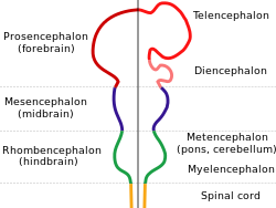
At the five-vesicle stage, the forebrain separates into the diencephalon (thalamus, hypothalamus, subthalamus, and epithalamus) and the telencephalon which develops into the cerebrum. The cerebrum consists of the cerebral cortex, underlying white matter, and the basal ganglia.

In humans, by 5 weeks in utero it is visible as a single portion toward the front of the fetus. At 8 weeks in utero, the forebrain splits into the left and right cerebral hemispheres.

When the embryonic forebrain fails to divide the brain into two lobes, it results in a condition known as holoprosencephaly. The parts present in the forebain are cerebrum, thalamus and hypothalamus

See also

Related Research Articles

<span class="mw-page-title-main">Brain</span> Organ that controls the nervous system in vertebrates and most invertebrates

The brain is an organ that serves as the center of the nervous system in all vertebrate and most invertebrate animals. In vertebrates, a small part of the brain called the hypothalamus is the neural control center for all endocrine systems. The brain is the largest cluster of neurons in the body and is typically located in the head, usually near organs for special senses such as vision, hearing and olfaction. It is the most energy-consuming organ of the body, and the most specialized, responsible for endocrine regulation, sensory perception, motor control, and the development of intelligence.

<span class="mw-page-title-main">Central nervous system</span> Brain and spinal cord

The central nervous system (CNS) is the part of the nervous system consisting of the brain and spinal cord, the retina and optic nerve, and the olfactory nerve and epithelia. The CNS is so named because the brain integrates the received information and coordinates and influences the activity of all parts of the bodies of bilaterally symmetric and triploblastic animals—that is, all multicellular animals except sponges and diploblasts. It is a structure composed of nervous tissue positioned along the rostral to caudal axis of the body and may have an enlarged section at the rostral end which is a brain. Only arthropods, cephalopods and vertebrates have a true brain, though precursor structures exist in onychophorans, gastropods and lancelets.

<span class="mw-page-title-main">Cerebral cortex</span> Outer layer of the cerebrum of the mammalian brain

The cerebral cortex, also known as the cerebral mantle, is the outer layer of neural tissue of the cerebrum of the brain in humans and other mammals. The cerebral cortex mostly consists of the six-layered neocortex, with just 10% consisting of the allocortex. It is separated into two cortices, by the longitudinal fissure that divides the cerebrum into the left and right cerebral hemispheres. The two hemispheres are joined beneath the cortex by the corpus callosum. The cerebral cortex is the largest site of neural integration in the central nervous system. It plays a key role in attention, perception, awareness, thought, memory, language, and consciousness. The cerebral cortex is part of the brain responsible for cognition.

<span class="mw-page-title-main">Thalamus</span> Structure within the brain

The thalamus is a large mass of gray matter located in the dorsal part of the diencephalon. Nerve fibers project out of the thalamus to the cerebral cortex in all directions, known as the thalamocortical radiations, allowing hub-like exchanges of information. It has several functions, such as the relaying of sensory signals, including motor signals to the cerebral cortex and the regulation of consciousness, sleep, and alertness.

<span class="mw-page-title-main">Holoprosencephaly</span> Medical condition

Holoprosencephaly (HPE) is a cephalic disorder in which the prosencephalon fails to develop into two hemispheres, typically occurring between the 18th and 28th day of gestation. Normally, the forebrain is formed and the face begins to develop in the fifth and sixth weeks of human pregnancy. The condition also occurs in other species.

<span class="mw-page-title-main">Brainstem</span> Posterior part of the brain, adjoining and structurally continuous

The brainstem is the stalk-like part of the brain that interconnects the cerebrum and diencephalon with the spinal cord. In the human brain, the brainstem is composed of the midbrain, the pons, and the medulla oblongata. The midbrain is continuous with the thalamus of the diencephalon through the tentorial notch.

<span class="mw-page-title-main">Ventricular system</span> Set of structures containing cerebrospinal fluid in the brain

In neuroanatomy, the ventricular system is a set of four interconnected cavities known as cerebral ventricles in the brain. Within each ventricle is a region of choroid plexus which produces the circulating cerebrospinal fluid (CSF). The ventricular system is continuous with the central canal of the spinal cord from the fourth ventricle, allowing for the flow of CSF to circulate.

<span class="mw-page-title-main">Third ventricle</span> Ventricle of the brain located between the two thalami

The third ventricle is one of the four connected ventricles of the ventricular system within the mammalian brain. It is a slit-like cavity formed in the diencephalon between the two thalami, in the midline between the right and left lateral ventricles, and is filled with cerebrospinal fluid (CSF).

<span class="mw-page-title-main">Midbrain</span> Forward-most portion of the brainstem

The midbrain or mesencephalon is the rostral-most portion of the brainstem connecting the diencephalon and cerebrum with the pons. It consists of the cerebral peduncles, tegmentum, and tectum.

<span class="mw-page-title-main">Cerebrum</span> Large part of the brain containing the cerebral cortex

The cerebrum, telencephalon or endbrain is the largest part of the brain containing the cerebral cortex, as well as several subcortical structures, including the hippocampus, basal ganglia, and olfactory bulb. In the human brain, the cerebrum is the uppermost region of the central nervous system. The cerebrum develops prenatally from the forebrain (prosencephalon). In mammals, the dorsal telencephalon, or pallium, develops into the cerebral cortex, and the ventral telencephalon, or subpallium, becomes the basal ganglia. The cerebrum is also divided into approximately symmetric left and right cerebral hemispheres.

<span class="mw-page-title-main">Human brain</span> Central organ of the human nervous system

The brain is the central organ of the human nervous system, and with the spinal cord makes up the central nervous system. The brain consists of the cerebrum, the brainstem and the cerebellum. It controls most of the activities of the body, processing, integrating, and coordinating the information it receives from the sense organs, and making decisions as to the instructions sent to the rest of the body. The brain is contained in, and protected by, the skull bones of the head.

<span class="mw-page-title-main">Diencephalon</span> Division of the forebrain around the third ventricle

In the human brain, the diencephalon is a division of the forebrain. It is situated between the telencephalon and the midbrain. The diencephalon has also been known as the tweenbrain in older literature. It consists of structures that are on either side of the third ventricle, including the thalamus, the hypothalamus, the epithalamus and the subthalamus.

<span class="mw-page-title-main">Reticular formation</span> Spinal trigeminal nucleus

The reticular formation is a set of interconnected nuclei that are located throughout the brainstem. It is not anatomically well defined, because it includes neurons located in different parts of the brain. The neurons of the reticular formation make up a complex set of networks in the core of the brainstem that extend from the upper part of the midbrain to the lower part of the medulla oblongata. The reticular formation includes ascending pathways to the cortex in the ascending reticular activating system (ARAS) and descending pathways to the spinal cord via the reticulospinal tracts.

The amygdalofugal pathway is one of the three major efferent pathways of the amygdala, meaning that it is one of the three principal pathways by which fibers leave the amygdala. It leads from the basolateral nucleus and central nucleus of the amygdala. The amygdala is a limbic structure in the medial temporal lobe of the brain. The other main efferent pathways from the amygdala are the stria terminalis and anterior commissure.

<span class="mw-page-title-main">Hypothalamic sulcus</span>

The hypothalamic sulcus is a groove in the lateral wall of the third ventricle, marking the boundary between the thalamus and hypothalamus. The upper and lower portions of the lateral wall of the third ventricle correspond to the alar lamina and basal lamina, respectively, of the lateral wall of the fore-brain vesicle and are separated from each other by a furrow, the hypothalamic sulcus, which extends from the interventricular foramen to the cerebral aqueduct.

The development of the nervous system in humans, or neural development or neurodevelopment involves the studies of embryology, developmental biology, and neuroscience to describe the cellular and molecular mechanisms by which the complex nervous system forms in humans, develops during prenatal development, and continues to develop postnatally.

Sleep onset is the transition from wakefulness into sleep. Sleep onset usually transmits into non-rapid eye movement sleep but under certain circumstances it is possible to transit from wakefulness directly into rapid eye movement sleep.

Dopaminergic cell groups, DA cell groups, or dopaminergic nuclei are collections of neurons in the central nervous system that synthesize the neurotransmitter dopamine. In the 1960s, dopaminergic neurons or dopamine neurons were first identified and named by Annica Dahlström and Kjell Fuxe, who used histochemical fluorescence. The subsequent discovery of genes encoding enzymes that synthesize dopamine, and transporters that incorporate dopamine into synaptic vesicles or reclaim it after synaptic release, enabled scientists to identify dopaminergic neurons by labeling gene or protein expression that is specific to these neurons.

<span class="mw-page-title-main">Brain vesicle</span>

Brain vesicles are the bulge-like enlargements of the early development of the neural tube in vertebrates, which eventually give rise to the brain.

References