Related Research Articles

In vertebrate anatomy, the pituitary gland is an endocrine gland. In humans, it is about the size of a chickpea and weighs, on average, 0.5 grams (0.018 oz). It is a protrusion off the bottom of the hypothalamus at the base of the brain. The hypophysis rests upon the hypophyseal fossa of the sphenoid bone in the center of the middle cranial fossa and is surrounded by a small bony cavity covered by a dural fold.

The hypothalamus is a part of the brain that contains a number of small nuclei with a variety of functions. One of the most important functions is to link the nervous system to the endocrine system via the pituitary gland. The hypothalamus is located below the thalamus and is part of the limbic system. In the terminology of neuroanatomy, it forms the ventral part of the diencephalon. All vertebrate brains contain a hypothalamus. In humans, it is the size of an almond.

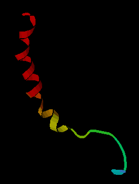
Human vasopressin, also called antidiuretic hormone (ADH), arginine vasopressin (AVP) or argipressin, is a hormone synthesized from the AVP gene as a peptide prohormone in neurons in the hypothalamus, and is converted to AVP. It then travels down the axon terminating in the posterior pituitary, and is released from vesicles into the circulation in response to extracellular fluid hypertonicity (hyperosmolality). AVP has two primary functions. First, it increases the amount of solute-free water reabsorbed back into the circulation from the filtrate in the kidney tubules of the nephrons. Second, AVP constricts arterioles, which increases peripheral vascular resistance and raises arterial blood pressure.

Oxytocin is a peptide hormone and neuropeptide normally produced in the hypothalamus and released by the posterior pituitary. Present in animals since early stages of evolution, in humans it plays roles in behavior that include social bonding, reproduction, childbirth, and the period after childbirth. Oxytocin is released into the bloodstream as a hormone in response to sexual activity and during labour. It is also available in pharmaceutical form. In either form, oxytocin stimulates uterine contractions to speed up the process of childbirth. In its natural form, it also plays a role in maternal bonding and milk production. Production and secretion of oxytocin is controlled by a positive feedback mechanism, where its initial release stimulates production and release of further oxytocin. For example, when oxytocin is released during a contraction of the uterus at the start of childbirth, this stimulates production and release of more oxytocin and an increase in the intensity and frequency of contractions. This process compounds in intensity and frequency and continues until the triggering activity ceases. A similar process takes place during lactation and during sexual activity.
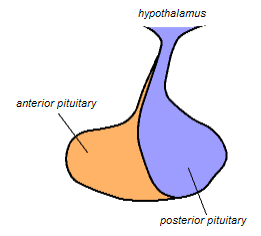
The posterior pituitary is the posterior lobe of the pituitary gland which is part of the endocrine system. The posterior pituitary is not glandular as is the anterior pituitary. Instead, it is largely a collection of axonal projections from the hypothalamus that terminate behind the anterior pituitary, and serve as a site for the secretion of neurohypophysial hormones directly into the blood. The hypothalamic–neurohypophyseal system is composed of the hypothalamus, posterior pituitary, and these axonal projections.

The supraoptic nucleus (SON) is a nucleus of magnocellular neurosecretory cells in the hypothalamus of the mammalian brain. The nucleus is situated at the base of the brain, adjacent to the optic chiasm. In humans, the SON contains about 3,000 neurons.

The paraventricular nucleus is a nucleus in the hypothalamus. Anatomically, it is adjacent to the third ventricle and many of its neurons project to the posterior pituitary. These projecting neurons secrete oxytocin and a smaller amount of vasopressin, otherwise the nucleus also secretes corticotropin-releasing hormone (CRH) and thyrotropin-releasing hormone (TRH). CRH and TRH are secreted into the hypophyseal portal system and act on different targets neurons in the anterior pituitary. PVN is thought to mediate many diverse functions through these different hormones, including osmoregulation, appetite, and the response of the body to stress.
Neuropeptides are chemical messengers made up of small chains of amino acids that are synthesized and released by neurons. Neuropeptides typically bind to G protein-coupled receptors (GPCRs) to modulate neural activity and other tissues like the gut, muscles, and heart.
A neurohormone is any hormone produced and released by neuroendocrine cells into the blood. By definition of being hormones, they are secreted into the circulation for systemic effect, but they can also have a role of neurotransmitter or other roles such as autocrine (self) or paracrine (local) messenger.
Neuroendocrine cells are cells that receive neuronal input and, as a consequence of this input, release messenger molecules (hormones) into the blood. In this way they bring about an integration between the nervous system and the endocrine system, a process known as neuroendocrine integration. An example of a neuroendocrine cell is a cell of the adrenal medulla, which releases adrenaline to the blood. The adrenal medullary cells are controlled by the sympathetic division of the autonomic nervous system. These cells are modified postganglionic neurons. Autonomic nerve fibers lead directly to them from the central nervous system. The adrenal medullary hormones are kept in vesicles much in the same way neurotransmitters are kept in neuronal vesicles. Hormonal effects can last up to ten times longer than those of neurotransmitters. Sympathetic nerve fiber impulses stimulate the release of adrenal medullary hormones. In this way the sympathetic division of the autonomic nervous system and the medullary secretions function together.
An osmoreceptor is a sensory receptor primarily found in the hypothalamus of most homeothermic organisms that detects changes in osmotic pressure. Osmoreceptors can be found in several structures, including two of the circumventricular organs – the vascular organ of the lamina terminalis, and the subfornical organ. They contribute to osmoregulation, controlling fluid balance in the body. Osmoreceptors are also found in the kidneys where they also modulate osmolality.
Neurophysin I is a carrier protein with a size of 10 KDa and contains 90 to 97 amino acids. It is a cleavage product of preprooxyphysin. It is a neurohypophysial hormone that is transported in vesicles with oxytocin, the other cleavage product, along axons, from magnocellular neurons of the hypothalamus to the posterior lobe of the pituitary. Although it is stored in neurosecretory granules with oxytocin and released with oxytocin, its biological action is unclear.

The subfornical organ (SFO) is one of the circumventricular organs of the brain. Its name comes from its location on the ventral surface of the fornix near the interventricular foramina, which interconnect the lateral ventricles and the third ventricle. Like all circumventricular organs, the subfornical organ is well-vascularized, and like all circumventricular organs except the subcommissural organ, some SFO capillaries have fenestrations, which increase capillary permeability. The SFO is considered a sensory circumventricular organ because it is responsive to a wide variety of hormones and neurotransmitters, as opposed to secretory circumventricular organs, which are specialized in the release of certain substances.

The pituitary stalk is the connection between the hypothalamus and the posterior pituitary. The floor of the third ventricle is prolonged downward as a funnel-shaped recess—the infundibular recess—into the infundibulum, where the apex of the pituitary is attached. It passes through the dura mater of the diaphragma sellae as it carries axons from the magnocellular neurosecretory cells of the hypothalamus down to the posterior pituitary where they release their neurohypophysial hormones, oxytocin and vasopressin, into the blood.
Neuroendocrinology is the branch of biology which studies the interaction between the nervous system and the endocrine system; i.e. how the brain regulates the hormonal activity in the body. The nervous and endocrine systems often act together in a process called neuroendocrine integration, to regulate the physiological processes of the human body. Neuroendocrinology arose from the recognition that the brain, especially the hypothalamus, controls secretion of pituitary gland hormones, and has subsequently expanded to investigate numerous interconnections of the endocrine and nervous systems.

In the nervous system, a synapse is a structure that permits a neuron to pass an electrical or chemical signal to another neuron or to the target effector cell.

Neurophysin II is a carrier protein with a size of 19,687.3 Da and is made up of a dimer of two virtually identical chains of amino acids. Neurophysin II is a cleavage product of the AVP gene. It is a neurohypophysial hormone that is transported in vesicles with vasopressin, the other cleavage product, along axons, from magnocellular neurons of the hypothalamus to the posterior lobe of the pituitary. Although it is stored in neurosecretory granules with vasopressin and released with vasopressin into the bloodstream, its biological action is unclear. Neurophysin II is also known as a stimulator of prolactin secretion.
Parvocellular neurosecretory cells are small neurons that produce hypothalamic releasing and inhibiting hormones. The cell bodies of these neurons are located in various nuclei of the hypothalamus or in closely related areas of the basal brain, mainly in the medial zone of the hypothalamus. All or most of the axons of the parvocellular neurosecretory cells project to the median eminence, at the base of the brain, where their nerve terminals release the hypothalamic hormones. These hormones are then immediately absorbed into the blood vessels of the hypothalamo-pituitary portal system, which carry them to the anterior pituitary gland, where they regulate the secretion of hormones into the systemic circulation.
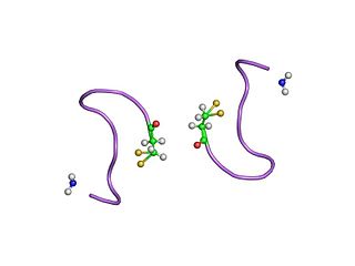
The neurohypophysial hormones form a family of structurally and functionally related peptide hormones. Their representatives in humans are oxytocin and vasopressin. They are named after the location of their release into the blood, the neurohypophysis.

Arginine Vasopressin (AVP) Gene is a gene whose product is proteolytically cleaved to produce vasopressin, neurophysin II, and a glycoprotein called copeptin. AVP and other AVP-like peptides are found in mammals, as well as mollusks, arthropods, nematodes, and other invertebrate species. In humans, AVP is present on chromosome 20 and plays a role in homeostatic regulation. The products of AVP have many functions that include vasoconstriction, regulating the balance of water in the body, and regulating responses to stress. Expression of AVP is regulated by the Transcription Translation Feedback Loop (TTFL), which is an important part of the circadian system that controls the expression of clock genes. AVP has important implications in the medical field as its products have significant roles throughout body.
References
- ↑ "BrainInfo". braininfo.rprc.washington.edu. Retrieved 16 August 2022.
- ↑ Leng, G; Brown, CH; Russell, JA (April 1999). "Physiological pathways regulating the activity of magnocellular neurosecretory cells". Progress in Neurobiology. 57 (6): 625–55. doi:10.1016/s0301-0082(98)00072-0. PMID 10221785. S2CID 240663.
- ↑ Ludwig, Mike; Leng, Gareth (31 January 2006). "Dendritic peptide release and peptide-dependent behaviours". Nature Reviews Neuroscience. 7 (2): 126–136. doi:10.1038/nrn1845. PMID 16429122. S2CID 31018227.
- ↑ Fisher, TE; Bourque, CW (Aug 1, 1995). "Voltage-gated calcium currents in the magnocellular neurosecretory cells of the rat supraoptic nucleus" (PDF). The Journal of Physiology. 486 (3): 571–80. doi:10.1113/jphysiol.1995.sp020835. PMC 1156547 . PMID 7473220.
- ↑ Ludwig, M; Sabatier, N; Dayanithi, G; Russell, JA; Leng, G (2002). The active role of dendrites in the regulation of magnocellular neurosecretory cell behavior. Progress in Brain Research. Vol. 139. pp. 247–56. doi:10.1016/s0079-6123(02)39021-6. ISBN 9780444509826. PMID 12436940.
- ↑ Brown, CH; Bains, JS; Ludwig, M; Stern, JE (August 2013). "Physiological regulation of magnocellular neurosecretory cell activity: integration of intrinsic, local and afferent mechanisms". Journal of Neuroendocrinology. 25 (8): 678–710. doi:10.1111/jne.12051. PMC 3852704 . PMID 23701531.