The thalamus is a large mass of gray matter in the dorsal part of the diencephalon of the brain with several functions such as relaying of sensory signals, including motor signals to the cerebral cortex, and the regulation of consciousness, sleep, and alertness.
Articles related to anatomy include:

The brainstem is the posterior part of the brain, continuous with the spinal cord. In the human brain the brainstem includes the midbrain, the pons and medulla oblongata of the hindbrain. The midbrain continues with the thalamus of the diencephalon through the tentorial notch, and sometimes the diencephalon is included in the brainstem.

The internal capsule is a white matter structure situated in the inferomedial part of each cerebral hemisphere of the brain. It carries information past the basal ganglia, separating the caudate nucleus and the thalamus from the putamen and the globus pallidus. The internal capsule contains both ascending and descending axons, going to and coming from the cerebral cortex. It also separates the caudate nucleus and the putamen in the dorsal striatum, a brain region involved in motor and reward pathways.

The grey column refers to a somewhat ridge-shaped mass of grey matter in the spinal cord. This presents as three columns: the anterior grey column, the posterior grey column, and the lateral grey column, all of which are visible in cross-section of the spinal cord.

The spinothalamic tract is a sensory pathway from the skin to the thalamus. From the ventral posterolateral nucleus in the thalamus, sensory information is relayed upward to the somatosensory cortex of the postcentral gyrus.

The thalamic reticular nucleus is part of the ventral thalamus that forms a capsule around the thalamus laterally. However, recent evidence from mice and fish question this statement and define it as dorsal thalamic structure. It is separated from the thalamus by the external medullary lamina. Reticular cells are GABAergic, and have discoid dendritic arbors in the plane of the nucleus.
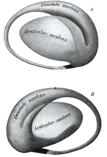
The lentiform nucleus or lenticular nucleus comprises the putamen and the globus pallidus within the basal ganglia. With the caudate nucleus it forms the striatum. It is a large, lens-shaped mass of gray matter just lateral to the internal capsule.

The spinocerebellar tract is a nerve tract originating in the spinal cord and terminating in the same side (ipsilateral) of the cerebellum.

The dentate nucleus is a cluster of neurons, or nerve cells, in the central nervous system that has a dentate – tooth-like or serrated – edge. It is located within the deep white matter of each cerebellar hemisphere, and it is the largest single structure linking the cerebellum to the rest of the brain. It is the largest and most lateral, or farthest from the midline, of the four pairs of deep cerebellar nuclei, the others being the fastigial nucleus and the globose and emboliform nuclei which together are referred to as the interposed nucleus. The dentate nucleus is responsible for the planning, initiation and control of voluntary movements. The dorsal region of the dentate nucleus contains output channels involved in motor function, which is the movement of skeletal muscle, while the ventral region contains output channels involved in nonmotor function, such as conscious thought and visuospatial function.

The habenular nuclei acts as a regulator of key central nervous system neurotransmitters, connecting the forebrain and midbrain within the epithalamus. Although it was predominantly studied for its demonstration of asymmetrical brain development and function, in recent years many scientists have begun to examine the habenular nuclei's role in motivation and behavior as it relates to an understanding of the physiology of addiction.

The septal area is an area in the lower, posterior part of the medial surface of the frontal lobe, and refers to the nearby septum pellucidum.

The substantia innominata also innominate substance, or substantia innominata of Meynert is a series of layers in the human brain consisting partly of gray and partly of white matter, which lies below the anterior part of the thalamus and lentiform nucleus. It is included as part of the anterior perforated substance. It is part of the basal forebrain structures and includes the nucleus basalis. A portion of the substantia innominata, below the globus pallidus is considered as part of the extended amygdala.

The anterior external arcuate fibers vary as to their prominence: in some cases they form an almost continuous layer covering the medullary pyramids and olivary body, while in other cases they are barely visible on the surface.
The mammillothalamic tract arises from cells in both the medial and lateral nuclei of the mammillary body and by fibers that are directly continued from the fornix.

The ansa lenticularis is a part of the brain, making up the superior layer of the substantia innominata. Its fibers, derived from the medullary lamina of the lentiform nucleus, pass medially to end in the thalamus and subthalamic region, while others are said to end in the tegmentum and red nucleus. It is classified by NeuroNames as part of the subthalamus.
The isothalamus is a division used by some researchers in describing the thalamus.

The stria medullaris is a part of the epithalamus. It is a fiber bundle containing afferent fibers from the septal nuclei, lateral preoptico-hypothalamic region, and anterior thalamic nuclei to the habenula. It forms a horizontal ridge on the medial surface of the thalamus, and is found on the border between dorsal and medial surfaces of thalamus. Superior and lateral to habenular trigone.

The spinal cord is a long, thin, tubular structure made up of nervous tissue, which extends from the medulla oblongata in the brainstem to the lumbar region of the vertebral column. It encloses the central canal of the spinal cord, which contains cerebrospinal fluid. The brain and spinal cord together make up the central nervous system (CNS). In humans, the spinal cord begins at the occipital bone, passing through the foramen magnum and entering the spinal canal at the beginning of the cervical vertebrae. The spinal cord extends down to between the first and second lumbar vertebrae, where it ends. The enclosing bony vertebral column protects the relatively shorter spinal cord. It is around 45 cm (18 in) in men and around 43 cm (17 in) long in women. The diameter of the spinal cord ranges from 13 mm in the cervical and lumbar regions to 6.4 mm in the thoracic area.