
The thalamus is a large mass of gray matter on the lateral walls of the third ventricle forming the dorsal part of the diencephalon. Nerve fibers project out of the thalamus to the cerebral cortex in all directions, known as the thalamocortical radiations, allowing hub-like exchanges of information. It has several functions, such as the relaying of sensory and motor signals to the cerebral cortex and the regulation of consciousness, sleep, and alertness.

The brainstem is the posterior stalk-like part of the brain that connects the cerebrum with the spinal cord. In the human brain the brainstem is composed of the midbrain, the pons, and the medulla oblongata. The midbrain is continuous with the thalamus of the diencephalon through the tentorial notch, and sometimes the diencephalon is included in the brainstem.

The internal capsule is a paired white matter structure, as a two-way tract, carrying ascending and descending fibers, to and from the cerebral cortex. The internal capsule is situated in the inferomedial part of each cerebral hemisphere of the brain. It carries information past the subcortical basal ganglia. As it courses it separates the caudate nucleus and the thalamus from the putamen and the globus pallidus. It also separates the caudate nucleus and the putamen in the dorsal striatum, a brain region involved in motor and reward pathways.

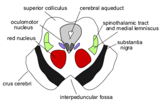
The spinothalamic tract is a nerve tract in the anterolateral system in the spinal cord. This tract is an ascending sensory pathway to the thalamus. From the ventral posterolateral nucleus in the thalamus, sensory information is relayed upward to the somatosensory cortex of the postcentral gyrus.

The pulvinar nuclei or nuclei of the pulvinar are the nuclei located in the thalamus. As a group they make up the collection called the pulvinar of the thalamus, usually just called the pulvinar.

The Papez circuit, or medial limbic circuit, is a neural circuit for the control of emotional expression. In 1937, James Papez proposed that the circuit connecting the hypothalamus to the limbic lobe was the basis for emotional experiences. Paul D. MacLean reconceptualized Papez's proposal and coined the term limbic system. MacLean redefined the circuit as the "visceral brain" which consisted of the limbic lobe and its major connections in the forebrain – hypothalamus, amygdala, and septum. Over time, the concept of a forebrain circuit for the control of emotional expression has been modified to include the prefrontal cortex.

The lateral corticospinal tract is the largest part of the corticospinal tract. It extends throughout the entire length of the spinal cord, and on transverse section appears as an oval area in front of the posterior column and medial to the posterior spinocerebellar tract.
The amygdalofugal pathway is one of the three major efferent pathways of the amygdala, meaning that it is one of the three principal pathways by which fibers leave the amygdala. It leads from the basolateral nucleus and central nucleus of the amygdala. The amygdala is a limbic structure in the medial temporal lobe of the brain. The other main efferent pathways from the amygdala are the stria terminalis and anterior commissure.
The mammillothalamic tract (MMT) is an efferent pathway of the mammillary bodies which project to the anterior nuclei of the thalamus. The mammillothalamic tract is part of the Papez circuit, starting and finishing in the hippocampus. The fibers of the MMT are heavily myelinated.

The medial dorsal nucleus is a large nucleus in the thalamus. It is separated from the other thalamic nuclei by the internal medullary lamina.
The anterior nuclei of thalamus are a collection of nuclei at the rostral end of the dorsal thalamus. They comprise the anteromedial, anterodorsal, and anteroventral nuclei.
The ventral posteromedial nucleus (VPM) is a nucleus of the thalamus and serves an analogous somatosensory relay role for the ascending trigeminothalamic tracts as its lateral neighbour the ventral posterolateral nucleus serves for dorsal column–medial lemniscus pathway 2nd-order neurons.

The intralaminar thalamic nuclei (ITN) are collections of neurons in the internal medullary lamina of the thalamus.
The trisynaptic circuit or trisynaptic loop is a relay of synaptic transmission in the hippocampus. The trisynaptic circuit is a neural circuit in the hippocampus, which is made up of three major cell groups: granule cells in the dentate gyrus, pyramidal neurons in CA3, and pyramidal neurons in CA1. The hippocampal relay involves 3 main regions within the hippocampus which are classified according to their cell type and projection fibers. The first projection of the hippocampus occurs between the entorhinal cortex (EC) and the dentate gyrus (DG). The entorhinal cortex transmits its signals from the parahippocampal gyrus to the dentate gyrus via granule cell fibers known collectively as the perforant path. The dentate gyrus then synapses on pyramidal cells in CA3 via mossy cell fibers. CA3 then fires to CA1 via Schaffer collaterals which synapse in the subiculum and are carried out through the fornix. Collectively the dentate gyrus, CA1 and CA3 of the hippocampus compose the trisynaptic loop.
The central tegmental tract is a structure in the midbrain and pons. It is situated in the central portion of the reticular formation. It contains:
The mammillotegmental fasciculus is a small bundle of efferent fibers from the hypothalamus running from the mammillary body to the tegmentum. Its functions are not well defined for humans, but based on animal studies it seems to be related to regulating visceral function and processing spatial information. The mammillotegmental fasciculus was first described by the German neuroanatomist, Bernhard von Gudden, from which it takes its alternate name, mammillo-tegmental bundle of Gudden.
The dorsal tegmental nucleus (DTN), also known as dorsal tegmental nucleus of Gudden (DTg), is a group of neurons located in the brain stem, which are involved in spatial navigation and orientation.
Patient N.A. was an American man who developed anterograde amnesia as a result of a fencing accident. He was a patient studied by Larry Squire, a professor of psychiatry, neuroscience and psychology at the University of California. The cause of his amnesia was found to be a thalamic lesion extending to the hypothalamus. Damage to the temporal cortex was also found and thought to be a result of an exploratory surgery.


