Related Research Articles

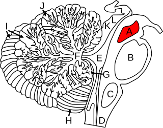
The brainstem is the posterior stalk-like part of the brain that connects the cerebrum with the spinal cord. In the human brain the brainstem is composed of the midbrain, the pons, and the medulla oblongata. The midbrain is continuous with the thalamus of the diencephalon through the tentorial notch, and sometimes the diencephalon is included in the brainstem.

The midbrain or mesencephalon is the rostral-most portion of the brainstem connecting the diencephalon and cerebrum with the pons. It consists of the cerebral peduncles, tegmentum, and tectum.

The mammillary bodies are a pair of small round bodies, located on the undersurface of the brain that, as part of the diencephalon, form part of the limbic system. They are located at the ends of the anterior arches of the fornix. They consist of two groups of nuclei, the medial mammillary nuclei and the lateral mammillary nuclei.

In neuroanatomy, a neural pathway is the connection formed by axons that project from neurons to make synapses onto neurons in another location, to enable neurotransmission. Neurons are connected by a single axon, or by a bundle of axons known as a nerve tract, or fasciculus. Shorter neural pathways are found within grey matter in the brain, whereas longer projections, made up of myelinated axons, constitute white matter.

The solitary nucleus(SN) (nucleus of the solitary tract, nucleus solitarius, or nucleus tractus solitarii) is a series of neurons whose cell bodies form a roughly vertical column of grey matter in the medulla oblongata of the brainstem. Their axons form the bulk of the enclosed solitary tract. The solitary nucleus can be divided into different parts including dorsomedial, dorsolateral, and ventrolateral subnuclei.

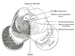
The medial longitudinal fasciculus (MLF) is a prominent bundle of nerve fibres which pass within the ventral/anterior portion of periaqueductal gray of the mesencephalon (midbrain). It contains the interstitial nucleus of Cajal, responsible for oculomotor control, head posture, and vertical eye movement.

The reticular formation is a set of interconnected nuclei that is located in the brainstem, hypothalamus, and other regions. It is not anatomically well defined, because it includes neurons located in different parts of the brain. The neurons of the reticular formation make up a complex set of networks in the core of the brainstem that extend from the upper part of the midbrain to the lower part of the medulla oblongata.

The pontine tegmentum, or dorsal pons, is the dorsal part of the pons located within the brainstem. The ventral part or ventral pons is known as the basilar part of the pons. The pontine tegmentum is all the material dorsal from the basilar pons to the fourth ventricle. Along with the dorsal surface of the medulla oblongata, it forms part of the rhomboid fossa – the floor of the fourth ventricle.

The spinocerebellar tracts are nerve tracts originating in the spinal cord and terminating in the same side (ipsilateral) of the cerebellum. The two main tracts are the dorsal spinocerebellar tract, and the ventral spinocerebellar tract. Both of these tracts are located in the peripheral region of the lateral funiculi.

The Papez circuit, or medial limbic circuit, is a neural circuit for the control of emotional expression. In 1937, James Papez proposed that the circuit connecting the hypothalamus to the limbic lobe was the basis for emotional experiences. Paul D. MacLean reconceptualized Papez's proposal and coined the term limbic system. MacLean redefined the circuit as the "visceral brain" which consisted of the limbic lobe and its major connections in the forebrain – hypothalamus, amygdala, and septum. Over time, the concept of a forebrain circuit for the control of emotional expression has been modified to include the prefrontal cortex.
The dorsal longitudinal fasciculus (DLF) is a longitudinal tract interconnecting the posterior hypothalamus, and the inferior medulla oblongata. It contains both ascending tracts and descending tracts, and serves to link the forebrain, and the visceral autonomic centres of the lower brainstem. It conveys both visceral motor signals, and sensory signals.
The mammillothalamic tract is an efferent pathway of the mammillary body which projects to the anterior nuclei of the thalamus. It consists of heavily myelinated fibres. It is part of a brain circuit involved in spatial memory.

The sensory decussation or decussation of the lemnisci is a decussation of axons from the gracile nucleus and cuneate nucleus, known together as the dorsal column nuclei. The dorsal column nuclei are responsible for fine touch, vibration, proprioception and two-point discrimination.

Johann Bernhard Aloys von Gudden was a German neuroanatomist and psychiatrist born in Kleve.

The basilar part of pons, also known as basis pontis, or basilar pons, is the ventral part of the pons in the brainstem; the dorsal part is known as the pontine tegmentum.
The midbrain reticular formation (MRF), also known as reticular formation of midbrain, mesencephalic reticular formation, tegmental reticular formation, and formatio reticularis (tegmenti) mesencephali, is a structure in the midbrain consisting of the dorsal tegmental nucleus, ventral tegmental nucleus, and cuneiform nucleus. These are also known as the tegmental nuclei.
The hypothalamospinal tract is an unmyelinated non-decussated descending nerve tract that arises in the hypothalamus and projects to the brainstem and spinal cord to synapse with pre-ganglionic autonomic neurons.
The interpeduncular nucleus (IPN) is an unpaired, ovoid cell group at the base of the midbrain tegmentum. It is located in the mesencephalon below the interpeduncular fossa. As the name suggests, the interpeduncular nucleus lies in between the cerebral peduncles.
The dorsal tegmental nucleus (DTN), also known as dorsal tegmental nucleus of Gudden (DTg), is a group of neurons located in the brain stem, which are involved in spatial navigation and orientation.
References
- ↑ Martin C. Hirsch (1999). Dictionary of Human Neuroanatomy. Springer. p. 79. ISBN 978-3540665236.
- ↑ Kwon, Hyeok Gyu; Hong, Ji Heon; Jang, Sung Ho (2011-05-03). "Mammillotegmental tract in the human brain: diffusion tensor tractography study". Neuroradiology. 53 (8): 623–626. doi:10.1007/s00234-011-0858-y. ISSN 0028-3940. PMID 21538047. S2CID 8494350.
- ↑ Donkelaar, Hans J. ten (2011-06-21). Clinical Neuroanatomy: Brain Circuitry and Its Disorders (1 ed.). Springer Berlin Heidelberg.
- ↑ Waxman, Stephen (2013-08-02). Clinical Neuroanatomy 27/E (27 ed.). New York: McGraw-Hill Medical. ISBN 9780071797979.