The brainstem is the posterior stalk-like part of the brain that connects the cerebrum with the spinal cord. In the human brain the brainstem is composed of the midbrain, the pons, and the medulla oblongata. The midbrain is continuous with the thalamus of the diencephalon through the tentorial notch, and sometimes the diencephalon is included in the brainstem.
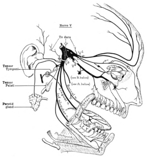
In neuroanatomy, the trigeminal nerve (lit. triplet nerve), also known as the fifth cranial nerve, cranial nerve V, or simply CN V, is a cranial nerve responsible for sensation in the face and motor functions such as biting and chewing; it is the most complex of the cranial nerves. Its name ("trigeminal", from Latin tri- 'three', and -geminus 'twin') derives from each of the two nerves (one on each side of the pons) having three major branches: the ophthalmic nerve (V1), the maxillary nerve (V2), and the mandibular nerve (V3). The ophthalmic and maxillary nerves are purely sensory, whereas the mandibular nerve supplies motor as well as sensory (or "cutaneous") functions. Adding to the complexity of this nerve is that autonomic nerve fibers as well as special sensory fibers (taste) are contained within it.

The glossopharyngeal nerve, also known as the ninth cranial nerve, cranial nerve IX, or simply CN IX, is a cranial nerve that exits the brainstem from the sides of the upper medulla, just anterior to the vagus nerve. Being a mixed nerve (sensorimotor), it carries afferent sensory and efferent motor information. The motor division of the glossopharyngeal nerve is derived from the basal plate of the embryonic medulla oblongata, whereas the sensory division originates from the cranial neural crest.

In neuroanatomy, the lateral geniculate nucleus is a structure in the thalamus and a key component of the mammalian visual pathway. It is a small, ovoid, ventral projection of the thalamus where the thalamus connects with the optic nerve. There are two LGNs, one on the left and another on the right side of the thalamus. In humans, both LGNs have six layers of neurons alternating with optic fibers.

The midbrain or mesencephalon is the forward-most portion of the brainstem and is associated with vision, hearing, motor control, sleep and wakefulness, arousal (alertness), and temperature regulation. The name comes from the Greek mesos, "middle", and enkephalos, "brain".

The spinothalamic tract is a part of the anterolateral system or the ventrolateral system, a sensory pathway to the thalamus. From the ventral posterolateral nucleus in the thalamus, sensory information is relayed upward to the somatosensory cortex of the postcentral gyrus.

The dorsal column–medial lemniscus pathway (DCML) is a sensory pathway of the central nervous system that conveys sensations of fine touch, vibration, two-point discrimination, and proprioception (position) from the skin and joints. It transmits information from the body to the primary somatosensory cortex in the postcentral gyrus of the parietal lobe of the brain. The pathway receives information from sensory receptors throughout the body, and carries this in nerve tracts in the white matter of the dorsal column of the spinal cord to the medulla, where it is continued in the medial lemniscus, on to the thalamus and relayed from there through the internal capsule and transmitted to the somatosensory cortex. The name dorsal-column medial lemniscus comes from the two structures that carry the sensory information: the dorsal columns of the spinal cord, and the medial lemniscus in the brainstem.
In neuroanatomy, thalamocortical radiations are the fibers between the thalamus and the cerebral cortex.

The dentate nucleus is a cluster of neurons, or nerve cells, in the central nervous system that has a dentate – tooth-like or serrated – edge. It is located within the deep white matter of each cerebellar hemisphere, and it is the largest single structure linking the cerebellum to the rest of the brain. It is the largest and most lateral, or farthest from the midline, of the four pairs of deep cerebellar nuclei, the others being the globose and emboliform nuclei, which together are referred to as the interposed nucleus, and the fastigial nucleus. The dentate nucleus is responsible for the planning, initiation and control of voluntary movements. The dorsal region of the dentate nucleus contains output channels involved in motor function, which is the movement of skeletal muscle, while the ventral region contains output channels involved in nonmotor function, such as conscious thought and visuospatial function.

The posterolateral tract is a small strand situated in relation to the tip of the posterior column close to the entrance of the posterior nerve roots. It is present throughout the spinal cord, and is most developed in the upper cervical regions.

In the human brain, the superior cerebellar peduncle is a paired structure of white matter that connects the cerebellum to the midbrain. It consists mainly of efferent fibers, the cerebellothalamic tract that runs from a cerebellar hemisphere to the contralateral thalamus, and the cerebellorubral tract that runs from a cerebellar hemisphere to the red nucleus. It also contains afferent tracts, most prominent of which is the ventral spinocerebellar tract. Other afferent tracts are the trigeminothalamic fibers, tectocerebellar fibers, and noradrenergic fibers from the locus coeruleus. The superior peduncle emerges from the upper and medial parts of the white matter of each hemisphere and is placed under cover of the upper part of the cerebellum.

The ventral posterior nucleus is the somato-sensory relay nucleus in thalamus of the brain.
The isothalamus is a division used by some researchers in describing the thalamus.
The principal sensory nucleus of trigeminal nerve is a group of second-order neurons which have cell bodies in the caudal pons.
The ventral posterolateral nucleus (VPL) is a nucleus of the thalamus. Together with the ventral posteromedial nucleus (VPM), ventral posterior inferior nucleus (VPI) and ventromedial posterior nucleus (VMpo), it constitutes the ventral posterior nucleus. There is uncertainty in the location of VMpo, as determined by spinothalamic tract (STT) terminations and staining for calcium-binding proteins, and several authorities do not consider its existence as being proved.
The ventrobasal complex (VB) is a relay nucleus of the thalamus for nociceptive stimuli received from nociceptive nerves. The VB consists of the ventral posteromedial nucleus (VPM) and the ventral posterolateral nucleus (VPL). In some species the ventral posterolateral nucleus, pars caudalis is also a part of the VB. The VB gets inputs from the spinothalamic tract, medial lemniscus, and corticothalamic tract. The main output of the VB is the primary somatosensory cortex.
The ventral trigeminal tract, ventral trigeminothalamic tract, anterior trigeminal tract, or anterior trigeminothalamic tract, is a tract composed of second order neuronal axons. These fibers carry sensory information about discriminative and crude touch, conscious proprioception, pain, and temperature from the head, face, and oral cavity. The ventral trigeminal tract connects the two major components of the brainstem trigeminal complex – the principal, or main sensory nucleus and the spinal trigeminal nucleus, to the ventral posteromedial nucleus of the thalamus.
The trigeminal lemniscus, also called the trigeminothalamic tract, is composed of the ventral trigeminal tract, and the dorsal trigeminal tract – nerve tracts that convey tactile, pain, and temperature impulses from the skin of the face, the mucous membranes of the nasal and oral cavities, and the eye, as well as proprioceptive information from the facial and masticatory muscles.
The dorsal trigeminal tract, dorsal trigeminothalamic tract, or posterior trigeminothalamic tract, is composed of second-order neuronal axons. These fibers carry sensory information about discriminative touch and conscious proprioception in the oral cavity from the principal nucleus of the trigeminal nerve to the ventral posteromedial (VPM) nucleus of the thalamus.
The central tegmental tract is a structure in the midbrain and pons.