Related Research Articles

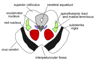
In humans, the tectospinal tract is a decussating extrapyramidal tract that coordinates head/neck and eye movements. It arises from the superior colliculus of the mesencephalic (midbrain) tectum, and projects to the cervical and upper thoracic spinal cord levels. It mediates reflex turning of the head and upper trunk in the direction of startling sensory stimuli.

The globose nucleus is one of the deep cerebellar nuclei. It is located medial to the emboliform nucleus, and lateral to the fastigial nucleus. The globose nucleus and emboliform nucleus are known collectively as the interposed nuclei.
The interposed nucleus is the combined globose and emboliform nuclei on either side. The interposed nucleus is one of the paired cerebellar nuclei. It is located in the roof of the fourth ventricle, lateral to the fastigial nucleus. The emboliform nucleus is the anterior interposed nucleus, and the globose nucleus is the posterior interposed nucleus.

The fastigial nucleus is located in each hemisphere of the cerebellum. It is one of the four deep cerebellar nuclei.

The flocculonodular lobe (vestibulocerebellum) is one of the lobes of the cerebellum. It is a small lobe consisting of the unpaired midline nodule and the two flocculi - one flocculus on either side of the nodule. The lobe is involved in maintaining posture and balance as well as coordinating head-eye movements.

The accessory cuneate nucleus is a nucleus situated in the caudal medulla oblongata just lateral to the cuneate nucleus. It relays unconscious proprioceptive sensory information from the upper limb and upper trunk to the cerebellum via the cuneocerebellar fibers.
The oral pontine reticular nucleus or rostral pontine reticular nucleus is one of the two components of the medial (efferent/motor) zone of the pontine reticular formation - the other being the caudal pontine reticular nucleus. The efferents of these two structures together give rise to the medial (pontine) reticulospinal tract. A population of their neurons together also form the paramedian pontine reticular formation which is involved in the coordination of horizontal conjugate eye movements in response to head movements.
The gigantocellular reticular nucleus is the (efferent/motor) medial zone of the reticular formation of the caudal pons and rostral medulla oblongata. It consists of a substantial number of giant neurons, but also contains small and medium sized neurons.
The spinoreticular tract is a partially decussating (crossed-over) four-neuron sensory pathway of the central nervous system. The tract transmits slow nociceptive/pain information from the spinal cord to reticular formation which in turn relays the information to the thalamus via reticulothalamic fibers as well as to other parts of the brain. Most (85%) second-order axons arising from sensory C first-order fibers ascend in the spinoreticular tract - it is consequently responsible for transmiting "slow", dull, poorly-localised pain. By projecting to the reticular activating system (RAS), the tract also mediates arousal/alertness in response to noxious (harmful) stimuli. The tract is phylogenetically older than the spinothalamic ("neospinothalamic") tract.

The juxtarestiform body is the smaller, medial subdivision of each inferior cerebellar peduncle.

The pontocerebellar fibers are the second-order neuron fibers of the corticopontocerebellar tracts that cross to the other side of the pons and run within the middle cerebellar peduncles, from the pons to the contralateral cerebellum. They arise from the pontine nuclei as the second part of the corticopontocerebellar tract, and decussate (cross-over) in the pons before passing through the middle cerebellar peduncles to reach and terminate in the contralateral posterior lobe of the cerebellum (neocerebellum). It is part of a pathway involved in the coordination of voluntary movements.
The dorsal trigeminal tract are uncrossed second-order sensory fibers conveying fine (discriminative) touch and pressure information from the dorsomedial division of principal sensory nucleus of trigeminal nerve to the ipsilateral ventral posteromedial nucleus of thalamus. Second-order fibers from the ventrolateral division of the principal sensory nucleus meanwhile cross-over to ascend contralaterally in the ventral trigeminal tract along with those fibers arising from the spinal trigeminal nucleus.

The central tegmental tract is a structure in the midbrain and pons.
The epidural venous plexus is a venous plexus embedded within the epidural fat of the vertebral canal. It is situated within the anterior epidural space. The plexus extends from the skull base to the sacrum. It is surrounded by sparse fat. It drains into the cavernous sinus of the cranial cavity; it also communicates with the radicular veins.
The hypothalamospinal tract is an unmyelinated non-decussated descending nerve tract that arises in the hypothalamus and projects to the brainstem and spinal cord to synapse with pre-ganglionic autonomic neurons.
The spinohypothalamic tract or spinohypothalamic fibers is a sensory fiber tract projecting from the spinal cord to the hypothalamus directly to mediate reflex autonomic and endocrine responses to painful stimuli (the hypothalamus receives additional indirect nociceptive projections from the reticular formation, and periaqueductal gray. The fibers of this tract synapse with hypothalamic neurons which in turn give rise to the hypothalamospinal tract that mediates the response of the autonomic nervous system to pain.
The cortico-olivary fibers are axons of neurons projecting from the primary motor cortex, premotor cortex, and somatosensory cortex bilaterally to both inferior olivary nuclei as part of the cortico-olivocerebellar pathway. They follows the same course as the corticopontine fibers. The inferior olivary nuclei subsequently project to the contralateral (cerebro)cerebellum via the olivocerebellar fibers. This pathway constitutes one of the three main afferent pathways of the cerebellum.
The nucleus of Darkschewitsch is an accessory oculomotor nucleus situated in the ventrolateral portion of the periaqueductal gray of the mesencephalon (midbrain) near its junction with the diencephalon. It is involved in mediating vertical eye movements. It projects to the trochlear nucleus, receives afferents from the visual cortex, and forms a reciprocal (looping) connection with the cerebellum by way of the inferior olive.
The nucleus of the posterior commissure is one of the accessory oculomotor nuclei situated in the mesencephalon (midbrain) at its junction with the diencephalon. It is involved in coordinating head-eye movements. It is situated near the oculomotor nucleus. It is thought to receive afferents from the ipsilateral cerebellum.

The spinomesencephalic pathway, spinomesencephalic tract or spino-quadrigeminal system of Mott, includes a number of ascending tracts in the spinal cord, including the spinotectal tract. The spinomesencephalic tract is one of the ascending tracts in the anterolateral system of the spinal cord that projects to various parts of the midbrain. It is involved in the processing of pain and visceral sensations.
References
- 1 2 Patestas, Maria A.; Gartner, Leslie P. (2016). A Textbook of Neuroanatomy (2nd ed.). Hoboken, New Jersey: Wiley-Blackwell. pp. 290–294. ISBN 978-1-118-67746-9.
- 1 2 Patestas, Maria A.; Gartner, Leslie P. (2016). A Textbook of Neuroanatomy (2nd ed.). Hoboken, New Jersey: Wiley-Blackwell. pp. 399–402. ISBN 978-1-118-67746-9.