Folium vermis

Last updated
Folium (vermis)

Gray702.png

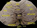
Upper surface of the cerebellum.
Identifiers
NeuroNames 675
TA A14.1.07.206
FMA 83889

Anatomical terms of neuroanatomy

The folium vermis is a short, narrow, concealed band at the posterior extremity of the vermis, consisting apparently of a single folium, but in reality marked on its upper and under surfaces by secondary fissures.

Cerebellar vermis Anatomical structure in the brain

The cerebellar vermis is located in the medial, cortico-nuclear zone of the cerebellum, which resides in the posterior fossa of the cranium. The primary fissure in the vermis curves ventrolaterally to the superior surface of the cerebellum, dividing it into anterior and posterior lobes. Functionally, the vermis is associated with bodily posture and locomotion. The vermis is included within the spinocerebellum and receives somatic sensory input from the head and proximal body parts via ascending spinal pathways.

Laterally, it expands in either hemisphere into a considerable lobule, the superior semilunar lobule (lobulus semilunaris superior; postero-superior lobules), which occupies the posterior third of the upper surface of the hemisphere, and is bounded below by the horizontal sulcus.

Horizontal fissure of cerebellum

The largest and deepest fissure in the cerebellum is named the horizontal fissure.

The superior semilunar lobules and the folium vermis form the lobus semilunaris.

Additional Images

Related Research Articles

Olecranon

The olecranon from the Greekolene meaning elbow and kranon meaning head is the large, thick, curved bony eminence of the ulna, a long bone in the forearm that projects behind the elbow. It forms the most pointed portion of the elbow and is opposite to the cubital fossa or elbow pit. The olecranon serves as a lever for the extensor muscles that straighten the elbow joint.

Flocculus (cerebellar)

The flocculus is a small lobe of the cerebellum at the posterior border of the middle cerebellar peduncle anterior to the biventer lobule. Like other parts of the cerebellum, the flocculus is involved in motor control. It is an essential part of the vestibulo-ocular reflex, and aids in the learning of basic motor skills in the brain.

Posterior inferior cerebellar artery

The posterior inferior cerebellar artery (PICA), the largest branch of the vertebral artery, is one of the three main arterial blood supplies for the cerebellum, part of the brain. Occlusion of the posterior inferior cerebellar artery or one of its branches, or of the vertebral artery leads to lateral medullary syndrome also called Wallenberg syndrome.

Semilunar hiatus

The semilunar hiatus or hiatus semilunaris, is a crescent-shaped groove in the lateral wall of the nasal cavity just inferior to the ethmoid bulla. It is the location of the openings for the frontal sinus, maxillary sinus, and anterior ethmoidal sinus. It is bounded inferiorly and anteriorly by the sharp concave margin of the uncinate process of the ethmoid bone, superiorly by the ethmoid bulla, and posteriorly by the middle nasal concha.

Squamous part of occipital bone

The squamous part of occipital bone, is situated above and behind the foramen magnum, and is curved from above downward and from side to side.

Cerebellar peduncles connect the cerebellum to the brain stem. There are six cerebellar peduncles in total, three on each side:

Lateral vestibular nucleus

The lateral vestibular nucleus is the continuation upward and lateralward of the principal nucleus, and in it terminate many of the ascending branches of the vestibular nerve.

Middle cerebellar peduncle

The middle cerebellar peduncles are paired structures that connect the cerebellum to the pons and are composed entirely of centripetal fibers, i.e. incoming fibers. The fibers arise from the pontine nucleus to the opposite hemisphere of the cerebellar cortex. The fibers are arranged in three fasciculi: superior, inferior, and deep.

Cerebellar veins

Cerebellar veins are veins which drain the cerebellum.

Lobe (anatomy) clear anatomical division or extension of an organ

In anatomy, a lobe is a clear anatomical division or extension of an organ that can be determined without the use of a microscope at the gross anatomy level. This is in contrast to the much smaller lobule, which is a clear division only visible under the microscope.

Cerebellar tonsil

The cerebellar tonsil is analogous to a rounded lobule on the undersurface of each cerebellar hemisphere, continuous medially with the uvula of the cerebellar vermis and superiorly by the flocculonodular lobe. Synonyms include: tonsilla cerebelli, amygdala cerebelli, the latter of which is not to be confused with the cerebral tonsils or amygdala nuclei located deep within the medial temporal lobes of the cerebral cortex. The flocculonodular lobe of the cerebellum which can also be confused for the cerebellar tonsils, is one of three lobes that make up the overall composition of the cerebellum. The cerebellum consists of three anatomical and functional lobes: anterior lobe, posterior lobe, and flocculonodular lobe.

Inferior medullary velum

The inferior medullary velum is a thin layer of white substance, prolonged from the white center of the cerebellum, above and on either side of the nodule; it forms the infero-posterior part of the fourth ventricle.

Vallecula of cerebellum

On the superior surface of cerebellum, the vermis protrudes above the level of the hemispheres, but on the inferior surface it is sunk almost out of sight in the bottom of a deep depression between them; this depression is called the vallecula of the cerebellum, and lodges the posterior part of the medulla oblongata and the inferior vermis, which consists of the tuber vermis, pyramid, uvula and nodule.

Nodule of vermis

The nodule, or anterior end of the inferior vermis, abuts against the roof of the fourth ventricle, and can only be distinctly seen after the cerebellum has been separated from the medulla oblongata and pons.

Tuber of vermis

The tuber of vermis, the most posterior division of the inferior vermis, is of small size, and laterally spreads out into the large inferior semilunar lobules, which comprise at least two-thirds of the inferior surface of the hemisphere.

Biventer lobule

The biventer lobule is a region of the cerebellum. It is triangular in shape; its apex points backward, and is joined by the gray band to the pyramid.

Central lobule

The central lobule is a small square lobule, situated in the anterior cerebellar notch. It overlaps the lingula, from which it is separated by the precentral fissure; laterally, it extends along the upper and anterior part of each hemisphere, where it forms a wing-like prolongation (ala), on each side, as the alae of the central lobule or alae lobuli centralis.

Culmen (cerebellum) structure in the brain

The culmen is the portion of the anterior vermis adjacent to the primary fissure of cerebellum.

Cerebellar cognitive affective syndrome (CCAS), also called "Schmahmann's syndrome" is a condition that follows from lesions (damage) to the cerebellum of the brain. It refers to a constellation of deficits in the cognitive domains of executive function, spatial cognition, language, and affect resulting from damage to the cerebellum. Impairments of executive function include problems with planning, set-shifting, abstract reasoning, verbal fluency, and working memory, and there is often perseveration, distractibility and inattention. Language problems include dysprosodia, agrammatism and mild anomia. Deficits in spatial cognition produce visual–spatial disorganization and impaired visual–spatial memory. Personality changes manifest as blunting of affect or disinhibited and inappropriate behavior. These cognitive impairments result in an overall lowering of intellectual function. CCAS challenges the traditional view of the cerebellum being responsible solely for regulation of motor functions. It is now thought that the cerebellum is responsible for monitoring both motor and nonmotor functions. The nonmotor deficits described in CCAS are believed to be caused by dysfunction in cerebellar connections to the cerebral cortex and limbic system.

References

This article incorporates text in the public domain from page 790 of the 20th edition of Gray's Anatomy (1918)

The public domain consists of all the creative works to which no exclusive intellectual property rights apply. Those rights may have expired, been forfeited, expressly waived, or may be inapplicable.

<i>Grays Anatomy</i> English-language textbook of human anatomy

Gray's Anatomy is an English language textbook of human anatomy originally written by Henry Gray and illustrated by Henry Vandyke Carter. Earlier editions were called Anatomy: Descriptive and Surgical, Anatomy of the Human Body and Gray's Anatomy: Descriptive and Applied, but the book's name is commonly shortened to, and later editions are titled, Gray's Anatomy. The book is widely regarded as an extremely influential work on the subject, and has continued to be revised and republished from its initial publication in 1858 to the present day. The latest edition of the book, the 41st, was published in September 2015.