Golgi cell

Last updated
Golgi cell
Diagram of the Microanatomy of Human Cerebellar Cortex.svg
Microcircuitry of the cerebellum. Excitatory synapses are denoted by (+) and inhibitory synapses by (-).
MF: Mossy fiber.
DCN: Deep cerebellar nuclei.
IO: Inferior olive.
CF: Climbing fiber.
GC: Granule cell.
PF: Parallel fiber.
PC: Purkinje cell.
GgC: Golgi cell.
SC: Stellate cell.
BC: Basket cell.
Details
Location Granular layer of the cerebellum
Identifiers
NeuroLex ID nifext_129
Anatomical terms of neuroanatomy

In neuroscience, Golgi cells are the most abundant inhibitory interneurons found within the granular layer of the cerebellum. [1] Golgi cells can be found in the granular layer at various layers. [2] The Golgi cell is essential for controlling the activity of the granular layer. [3] They were first identified as inhibitory in 1964. [4] It was also the first example of an inhibitory feedback network in which the inhibitory interneuron was identified anatomically. Golgi cells produce a wide lateral inhibition that reaches beyond the afferent synaptic field and inhibit granule cells via feedforward and feedback inhibitory loops. [4] These cells synapse onto the dendrite of granule cells and unipolar brush cells. They receive excitatory input from mossy fibres, also synapsing on granule cells, and parallel fibers, which are long granule cell axons. Thereby this circuitry allows for feed-forward and feed-back inhibition of granule cells.

Contents

Connections

The cerebellar network contains a large number of connections between Golgi cells. [5] The main synapse made by these cells is a synapse onto the mossy fibre–granule cell excitatory synapse in a glomerulus. The glomerulus is made up of the mossy fibre terminal, granule cell dendrites, and the Golgi terminal, and is enclosed by a glial coat. [3] The Golgi cell acts by altering the mossy fibre - granule cell synapse. According to reports, granule cells mostly connect to the Golgi cell through parallel fibers, while synapses may also play a role. It has been shown that the climbing fibers connect to the Golgi cells by reentering the higher granular layer through thin collateral branches that reach slightly below the Purkinje cells. [6] Three different types of inhibitory interneurons—basket and stellate cells, which are found in the molecular layer, and Golgi cells, which are found in the granular layer—are triggered by parallel fibers and control the activity of Purkinje cells. [7]

In the theta frequency range, Golgi cells exhibit pacemaking, resonance, phase-reset, and rebound-excitation. These characteristics probably have an effect on their behavior. In vivo, exhibiting erratic, spontaneous beating regulated by sensory inputs and sudden, quiet pauses between burst responses to punctuate stimulus. Furthermore, the network's Golgi cell interaction offers insight into how these neurons may control the spatiotemporal arrangement of cerebellar activity. It turns out that Golgi cells can affect both the temporal dynamics and the geographical distribution of information relayed across the cerebellar network. Golgi cells also control the mossy fiber–granule cell synapse's production of long-term synaptic plasticity. Thus, the idea that Golgi cells play a crucial role in controlling the activity of the granular layer network, which has significant implications for cerebellar computing, is beginning to take shape. [8]

Glutamatergic stimuli are the primary excitatory inputs to Golgi cells. Current research indicates that NMDA [9] receptors and AMPA [10] receptors are involved at mossy fiber-Golgi cell relays. [11] Golgi cell circuit functions also seem to be regulated by metabotropic glutamate receptors. Golgi cells possess mGluR2 receptors, [12] and when these receptors are activated, an inward rectifier K current is enhanced, aiding in the Golgi cell's silencing after a period of intensive granule cell-Golgi cell transmission. [13] This mGluR2-dependent process could make it easier for extended bursts to travel through the mossy fiber-granule cell route. [14]

Neurotransmitters

Golgi cells predominantly use GABA and glycine as neurotransmitters, although depending on their target, a single Golgi cell will selectively facilitate GABAergic or glycinergic transmission. When Golgi cells are activated, granule cells and UBCs exhibit GABAergic and glycinergic currents, respectively. This is primarily because Golgi cells corelease GABA and glycine at each individual bouton, which is a result of cell-type-specific postsynaptic receptor expression and/or trafficking to the synapse. [15] Based on electron microscopy [16] and electrophysiological evidence, [17] it was believed that molecular layer interneurons (stellate and basket cells) form GABAergic synapses on Golgi cell dendrites. However, more recent research challenges the existence of these functional synapses. [18] Instead, local interneurons provide just a small fraction of the inhibitory inputs to Golgi cells in the molecular layer, and most of these inputs only release GABA. Rather, Golgi cells are innervated by GABAergic cells in the DCN, indicating that Golgi cells get a large input of feedback inhibition from the deep cerebellar nuclei to modulate local inhibitory networks. [19] The basal level of GABA produces a postsynaptic leak conductance by tonically activating alpha 6-containing GABA-A receptors on the granule cell. [1] [2] [10] These high-affinity receptors are located both synaptically and extrasynaptically on the granule cell. The synaptic receptors mediate phasic contraction, duration of around 20–30ms, whereas the extrasynapatic receptors mediate tonic inhibition of around 200ms, and are activated by synapse spill over. [9]

Additionally the GABA acts on GABA-B receptors which are located presynaptically on the mossy fibre terminal. These inhibit the mossy fibre evoked EPSCs of the granule cell in a temperature and frequency dependent manner. At high mossy firing frequency (10 Hz) there is no effect of GABA acting on presynaptic GABA-B receptors on evoked EPSCs. However, at low (1 Hz) firing the GABA does have an effect on the EPSCs mediated via these presynaptic GABA-B receptors.

Golgi cells are necessary for complex motor coordination, as this study shows "Ablation of Cerebellar Golgi Cells Disrupts Synaptic Integration Involving GABA Inhibition and NMDA Receptor Activation in Motor Coordination" conducted by Watanabe, D., Inokawa, H., et al. Moreover, such compound motions depend on a synaptic integration that is generated from granule cell NMDA receptor activation and GABA-mediated inhibition. [7] Eventually, the Golgi cells use an expanded axonal plexus to block broad fields of granule cells. The fundamental questions of whether dendritic processing underlies the theory-predicted spike-timing dependent plasticity (STDP) and how synaptic inputs govern the generation of Golgi cell spikes remain unanswered [20] . It's interesting to note that dendrites express a diverse set of Ca, Na, and K ionic channels [21] that may have an effect on dendritic computation, while mossy fiber–Golgi cell synapses express NMDA channels, which are essential for synaptic plasticity. [22] Making a forecast regarding the potential interconnections between these many active features is challenging and necessitates a thorough computational examination of synaptic integration and the neuron's electrogenic architecture. [23]

Golgi type I

The cell bodies of Golgi type I neurons are medium-to-large. [24] A Golgi type I neuron has a long axon that begins in the grey matter of the central nervous system and may extend from there. Their cell bodies were mostly multipolar, yet occasionally they might have been triangular in shape and lacking any appendages or spines. They possessed three to ten principal dendrites. There were no appendages or spines on these dendrites. The cells' dendrites had abundant arborizations. [25]

These neurons have tufted and radiated branching patterns in their dendrites compared to the tufted pattern, the radiating branching pattern was more prevalent. [26] The density of dendritic trees is typically present in these cells, but the quantity and diameter of primary dendrites are highly irregular. Outside the cell body, three to eleven dendrites are visible. Prior to splitting into tertiary branches, it quickly give rise to thinner secondary dendrites. [27]

It is also known as a projection neuron. They include the neurons forming peripheral nerves and long tracts of brain and spinal cord. [11] with somata usually ranging from 20 to 40µm. [27] Golgi II neurons, in contrast, are defined as having short axons or no axon at all. This distinction was introduced by the pioneering neuroanatomist Camillo Golgi, on the basis of the appearance under a microscope of neurons stained with the Golgi stain that he had invented. Santiago Ramón y Cajal postulated that higher developed animals had more Golgi type II in comparison to Golgi type I neurons. These Golgi type II neurons have a star-like appearance, and are found in cerebral and cerebellar cortices and retina. [28]

Golgi type II

These neurons' cell bodies were ovoid, spheroid, or multipolar. [27] A Golgi type II neuron either has no axon or else a short axon that does not send branches out of the gray matter of the central nervous system. [12] Golgi type II dendrites have approximately symmetrical synaptic connections and have pale, asymmetric, and frequently massive profiles that contain huge pleomorphic vesicles. Golgi type II axon synaptic terminals may resemble dendritic endings, however many axonal endings seem to have narrower profiles with smaller, flatter vesicles. [29] Their average diameter varied from 12 to 30 lm, with a mean of 22.2 lm on average (5.8 ± n = 120). [27] Compared to Golgi type I neurons, Golgi type II neurons have a greater nucleus to cytoplasm ratio (N/C). [27] Compared to Golgi type I neurons, these neurons' dendrites exhibit significantly less tufted dendrites. Two in the ten main dendrites protruded from the cell body and produced a small number of branches. [27] Golgi type II neuron generates dendro-dendritic connections with the main neuron in terminal aggregates termed synaptic nests. Afferent axons descending from the auditory cortex and ascending from the posterior colliculus form synaptic connections with both kinds of neurons. [30]

The Golgi type II cells might be excitatory or inhibitory interneurons, or they can be both. Golgi type II cells function as inhibitory interneurons, which could produce response patterns that make the primary neurons more responsive to the beginning of stimuli and to temporal variations in the afferent input. Golgi type II cells, being excitatory interneurons, have the ability to produce gradual or continuous response patterns that have the tendency to extend specific signal trains. In each scenario, the cortical analysis of sound locations and temporal patterns depends on the synaptic interactions between Golgi type II cells to define the spatial and temporal features of stimulus coding. [31]

See also

List of distinct cell types in the adult human body

Related Research Articles

<span class="mw-page-title-main">Neuron</span> Electrically excitable cell found in the nervous system of animals

Within a nervous system, a neuron, neurone, or nerve cell is an electrically excitable cell that fires electric signals called action potentials across a neural network. Neurons communicate with other cells via synapses, which are specialized connections that commonly use minute amounts of chemical neurotransmitters to pass the electric signal from the presynaptic neuron to the target cell through the synaptic gap.

<span class="mw-page-title-main">Cerebellum</span> Structure at the rear of the vertebrate brain, beneath the cerebrum

The cerebellum is a major feature of the hindbrain of all vertebrates. Although usually smaller than the cerebrum, in some animals such as the mormyrid fishes it may be as large as it or even larger. In humans, the cerebellum plays an important role in motor control. It may also be involved in some cognitive functions such as attention and language as well as emotional control such as regulating fear and pleasure responses, but its movement-related functions are the most solidly established. The human cerebellum does not initiate movement, but contributes to coordination, precision, and accurate timing: it receives input from sensory systems of the spinal cord and from other parts of the brain, and integrates these inputs to fine-tune motor activity. Cerebellar damage produces disorders in fine movement, equilibrium, posture, and motor learning in humans.

<span class="mw-page-title-main">Olfactory bulb</span> Neural structure

The olfactory bulb is a neural structure of the vertebrate forebrain involved in olfaction, the sense of smell. It sends olfactory information to be further processed in the amygdala, the orbitofrontal cortex (OFC) and the hippocampus where it plays a role in emotion, memory and learning. The bulb is divided into two distinct structures: the main olfactory bulb and the accessory olfactory bulb. The main olfactory bulb connects to the amygdala via the piriform cortex of the primary olfactory cortex and directly projects from the main olfactory bulb to specific amygdala areas. The accessory olfactory bulb resides on the dorsal-posterior region of the main olfactory bulb and forms a parallel pathway. Destruction of the olfactory bulb results in ipsilateral anosmia, while irritative lesions of the uncus can result in olfactory and gustatory hallucinations.

An inhibitory postsynaptic potential (IPSP) is a kind of synaptic potential that makes a postsynaptic neuron less likely to generate an action potential. The opposite of an inhibitory postsynaptic potential is an excitatory postsynaptic potential (EPSP), which is a synaptic potential that makes a postsynaptic neuron more likely to generate an action potential. IPSPs can take place at all chemical synapses, which use the secretion of neurotransmitters to create cell-to-cell signalling. EPSPs and IPSPs compete with each other at numerous synapses of a neuron. This determines whether an action potential occurring at the presynaptic terminal produces an action potential at the postsynaptic membrane. Some common neurotransmitters involved in IPSPs are GABA and glycine.

<span class="mw-page-title-main">Pyramidal cell</span> Projection neurons in the cerebral cortex and hippocampus

Pyramidal cells, or pyramidal neurons, are a type of multipolar neuron found in areas of the brain including the cerebral cortex, the hippocampus, and the amygdala. Pyramidal cells are the primary excitation units of the mammalian prefrontal cortex and the corticospinal tract. Pyramidal neurons are also one of two cell types where the characteristic sign, Negri bodies, are found in post-mortem rabies infection. Pyramidal neurons were first discovered and studied by Santiago Ramón y Cajal. Since then, studies on pyramidal neurons have focused on topics ranging from neuroplasticity to cognition.

<span class="mw-page-title-main">Basket cell</span>

Basket cells are inhibitory GABAergic interneurons of the brain, found throughout different regions of the cortex and cerebellum.

An apical dendrite is a dendrite that emerges from the apex of a pyramidal cell. Apical dendrites are one of two primary categories of dendrites, and they distinguish the pyramidal cells from spiny stellate cells in the cortices. Pyramidal cells are found in the prefrontal cortex, the hippocampus, the entorhinal cortex, the olfactory cortex, and other areas. Dendrite arbors formed by apical dendrites are the means by which synaptic inputs into a cell are integrated. The apical dendrites in these regions contribute significantly to memory, learning, and sensory associations by modulating the excitatory and inhibitory signals received by the pyramidal cells.

<span class="mw-page-title-main">Neurotransmission</span> Impulse transmission between neurons

Neurotransmission is the process by which signaling molecules called neurotransmitters are released by the axon terminal of a neuron, and bind to and react with the receptors on the dendrites of another neuron a short distance away. A similar process occurs in retrograde neurotransmission, where the dendrites of the postsynaptic neuron release retrograde neurotransmitters that signal through receptors that are located on the axon terminal of the presynaptic neuron, mainly at GABAergic and glutamatergic synapses.

Depolarization-induced suppression of inhibition is the classical and original electrophysiological example of endocannabinoid function in the central nervous system. Prior to the demonstration that depolarization-induced suppression of inhibition was dependent on the cannabinoid CB1 receptor function, there was no way of producing an in vitro endocannabinoid mediated effect.

The stratum lucidum of the hippocampus is a layer of the hippocampus between the stratum pyramidale and the stratum radiatum. It is the tract of the mossy fiber projections, both inhibitory and excitatory from the granule cells of the dentate gyrus. One mossy fiber may make up to 37 connections to a single pyramidal cell, and innervate around 12 pyramidal cells on top of that. Any given pyramidal cell in the stratum lucidum may get input from as many as 50 granule cells.

<span class="mw-page-title-main">Mossy fiber (hippocampus)</span> Pathway in the hippocampus

In the hippocampus, the mossy fiber pathway consists of unmyelinated axons projecting from granule cells in the dentate gyrus that terminate on modulatory hilar mossy cells and in Cornu Ammonis area 3 (CA3), a region involved in encoding short-term memory. These axons were first described as mossy fibers by Santiago Ramón y Cajal as they displayed varicosities along their lengths that gave them a mossy appearance. The axons that make up the pathway emerge from the basal portions of the granule cells and pass through the hilus of the dentate gyrus before entering the stratum lucidum of CA3. Granule cell synapses tend to be glutamatergic, though immunohistological data has indicated that some synapses contain neuropeptidergic elements including opiate peptides such as dynorphin and enkephalin. There is also evidence for co-localization of both GABAergic and glutamatergic neurotransmitters within mossy fiber terminals. GABAergic and glutamatergic co-localization in mossy fiber boutons has been observed primarily in the developing hippocampus, but in adulthood, evidence suggests that mossy fiber synapses may alternate which neurotransmitter is released through activity-dependent regulation.

<span class="mw-page-title-main">Hippocampus anatomy</span>

Hippocampus anatomy describes the physical aspects and properties of the hippocampus, a neural structure in the medial temporal lobe of the brain. It has a distinctive, curved shape that has been likened to the sea-horse monster of Greek mythology and the ram's horns of Amun in Egyptian mythology. This general layout holds across the full range of mammalian species, from hedgehog to human, although the details vary. For example, in the rat, the two hippocampi look similar to a pair of bananas, joined at the stems. In primate brains, including humans, the portion of the hippocampus near the base of the temporal lobe is much broader than the part at the top. Due to the three-dimensional curvature of this structure, two-dimensional sections such as shown are commonly seen. Neuroimaging pictures can show a number of different shapes, depending on the angle and location of the cut.

<span class="mw-page-title-main">Anatomy of the cerebellum</span> Structures in the cerebellum, a part of the brain

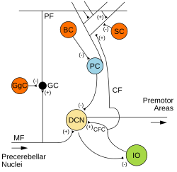
The anatomy of the cerebellum can be viewed at three levels. At the level of gross anatomy, the cerebellum consists of a tightly folded and crumpled layer of cortex, with white matter underneath, several deep nuclei embedded in the white matter, and a fluid-filled ventricle in the middle. At the intermediate level, the cerebellum and its auxiliary structures can be broken down into several hundred or thousand independently functioning modules or compartments known as microzones. At the microscopic level, each module consists of the same small set of neuronal elements, laid out with a highly stereotyped geometry.

<span class="mw-page-title-main">Cerebellar granule cell</span> Thick granular layer of the cerebellar cortex

Cerebellar granule cells form the thick granular layer of the cerebellar cortex and are among the smallest neurons in the brain. Cerebellar granule cells are also the most numerous neurons in the brain: in humans, estimates of their total number average around 50 billion, which means that they constitute about 3/4 of the brain's neurons.

<span class="mw-page-title-main">Granule cell</span> Type of neuron with a very small cell body

The name granule cell has been used for a number of different types of neurons whose only common feature is that they all have very small cell bodies. Granule cells are found within the granular layer of the cerebellum, the dentate gyrus of the hippocampus, the superficial layer of the dorsal cochlear nucleus, the olfactory bulb, and the cerebral cortex.

An autapse is a chemical or electrical synapse from a neuron onto itself. It can also be described as a synapse formed by the axon of a neuron on its own dendrites, in vivo or in vitro.

<span class="mw-page-title-main">Unipolar brush cell</span>

Unipolar brush cells (UBCs) are a class of excitatory glutamatergic interneuron found in the granular layer of the cerebellar cortex and also in the granule cell domain of the cochlear nucleus.

<span class="mw-page-title-main">Glomerulus (cerebellum)</span>

The cerebellar glomerulus is a small, intertwined mass of nerve fiber terminals in the granular layer of the cerebellar cortex. It consists of post-synaptic granule cell dendrites and pre-synaptic terminals of mossy fibers.

<span class="mw-page-title-main">Hippocampus proper</span> Part of the brain of mammals

The hippocampus proper refers to the actual structure of the hippocampus which is made up of three regions or subfields. The subfields CA1, CA2, and CA3 use the initials of cornu Ammonis, an earlier name of the hippocampus.

An axo-axonic synapse is a type of synapse, formed by one neuron projecting its axon terminals onto another neuron's axon.

References

  1. 1 2 Brickley SG, Cull-Candy SG, Farrant M (December 1996). "Development of a tonic form of synaptic inhibition in rat cerebellar granule cells resulting from persistent activation of GABAA receptors". The Journal of Physiology. 497 ( Pt 3) (Pt 3): 753–759. doi:10.1113/jphysiol.1996.sp021806. PMC   1160971 . PMID   9003560.
  2. 1 2 Tia S, Wang JF, Kotchabhakdi N, Vicini S (June 1996). "Developmental changes of inhibitory synaptic currents in cerebellar granule neurons: role of GABA(A) receptor alpha 6 subunit". The Journal of Neuroscience. 16 (11): 3630–3640. doi: 10.1523/JNEUROSCI.16-11-03630.1996 . PMC   6578841 . PMID   8642407.
  3. 1 2 Jakab RL, Hámori J (1988). "Quantitative morphology and synaptology of cerebellar glomeruli in the rat". Anatomy and Embryology. 179 (1): 81–88. doi:10.1007/BF00305102. PMID   3213958. S2CID   22651721.
  4. 1 2 Eccles J, Llinas R, Sasaki K (December 1964). "Golgi cell inhibition in the cerebellar cortex". Nature. 204 (4965): 1265–1266. Bibcode:1964Natur.204.1265E. doi:10.1038/2041265a0. PMID   14254404. S2CID   4187138.
  5. Palay SL, Chan-Palay V (1974). Cerebellar Cortex. doi:10.1007/978-3-642-65581-4. ISBN   978-3-642-65583-8. S2CID   31651518.
  6. Shinoda Y, Sugihara I, Wu HS, Sugiuchi Y (2000). "The entire trajectory of single climbing and mossy fibers in the cerebellar nuclei and cortex". Cerebellar modules: Molecules, morphology and function. Progress in Brain Research. Vol. 124. Elsevier. pp. 173–186. doi:10.1016/s0079-6123(00)24015-6. ISBN   978-0-444-50108-0. PMID   10943124.
  7. 1 2 Watanabe D, Inokawa H, Hashimoto K, Suzuki N, Kano M, Shigemoto R, et al. (October 1998). "Ablation of cerebellar Golgi cells disrupts synaptic integration involving GABA inhibition and NMDA receptor activation in motor coordination". Cell. 95 (1): 17–27. doi:10.1016/s0092-8674(00)81779-1. hdl: 2433/181271 . PMID   9778244.
  8. D'Angelo E (July 2008). "The critical role of Golgi cells in regulating spatio-temporal integration and plasticity at the cerebellum input stage". Frontiers in Neuroscience. 2 (1): 35–46. doi: 10.3389/neuro.01.008.2008 . PMC   2570065 . PMID   18982105.
  9. 1 2 Nusser Z, Sieghart W, Somogyi P (March 1998). "Segregation of different GABAA receptors to synaptic and extrasynaptic membranes of cerebellar granule cells" (abstract). The Journal of Neuroscience. 18 (5): 1693–1703. doi:10.1523/JNEUROSCI.18-05-01693.1998. PMC   6792611 . PMID   9464994.
  10. 1 2 Wall MJ, Usowicz MM (March 1997). "Development of action potential-dependent and independent spontaneous GABAA receptor-mediated currents in granule cells of postnatal rat cerebellum". The European Journal of Neuroscience. 9 (3): 533–548. doi:10.1111/j.1460-9568.1997.tb01630.x. PMID   9104595. S2CID   39603115.
  11. 1 2 "Golgi type I neuron definition". Dictionary.com. 2008. Retrieved 2008-12-25.
  12. 1 2 "Golgi type II neuron definition". Dictionary.com. Retrieved 2019-08-15.
  13. Watanabe D, Nakanishi S (August 2003). "mGluR2 postsynaptically senses granule cell inputs at Golgi cell synapses". Neuron. 39 (5): 821–829. doi: 10.1016/S0896-6273(03)00530-0 . PMID   12948448.
  14. Arenz A, Silver RA, Schaefer AT, Margrie TW (August 2008). "The contribution of single synapses to sensory representation in vivo". Science. 321 (5891): 977–980. Bibcode:2008Sci...321..977A. doi:10.1126/science.1158391. PMC   2771362 . PMID   18703744.
  15. Dugué GP, Dumoulin A, Triller A, Dieudonné S (July 2005). "Target-dependent use of co-released inhibitory transmitters at central synapses". The Journal of Neuroscience. 25 (28): 6490–6498. doi:10.1523/jneurosci.1500-05.2005. PMC   6725433 . PMID   16014710.
  16. Chan-Palay V, Palay SL, Billings-Gagliardi SM (November 1974). "Meynert cells in the primate visual cortex". Journal of Neurocytology. 3 (5): 631–658. doi:10.1007/bf01097628. PMID   4142639. S2CID   25165435.
  17. Dumoulin A, Triller A, Dieudonné S (August 2001). "IPSC kinetics at identified GABAergic and mixed GABAergic and glycinergic synapses onto cerebellar Golgi cells". The Journal of Neuroscience. 21 (16): 6045–6057. doi:10.1523/jneurosci.21-16-06045.2001. PMC   6763194 . PMID   11487628.
  18. Eyre MD, Nusser Z (May 2016). "Only a Minority of the Inhibitory Inputs to Cerebellar Golgi Cells Originates from Local GABAergic Cells". eNeuro. 3 (2): ENEURO.0055–16.2016. doi:10.1523/eneuro.0055-16.2016. PMC   4876488 . PMID   27257627.
  19. Ankri L, Husson Z, Pietrajtis K, Proville R, Léna C, Yarom Y, Dieudonné S, Uusisaari MY (2015-04-22). "Author response: A novel inhibitory nucleo-cortical circuit controls cerebellar Golgi cell activity". eLife. doi: 10.7554/elife.06262.015 .
  20. Garrido JA, Luque NR, Tolu S, D'Angelo E (August 2016). "Oscillation-Driven Spike-Timing Dependent Plasticity Allows Multiple Overlapping Pattern Recognition in Inhibitory Interneuron Networks". International Journal of Neural Systems. 26 (5): 1650020. doi:10.1142/S0129065716500209. PMID   27079422. S2CID   9779306.
  21. Rudolph S, Hull C, Regehr WG (November 2015). "Active Dendrites and Differential Distribution of Calcium Channels Enable Functional Compartmentalization of Golgi Cells". The Journal of Neuroscience. 35 (47): 15492–15504. doi:10.1523/JNEUROSCI.3132-15.2015. PMC   4659820 . PMID   26609148.
  22. Cesana E, Pietrajtis K, Bidoret C, Isope P, D'Angelo E, Dieudonné S, Forti L (July 2013). "Granule cell ascending axon excitatory synapses onto Golgi cells implement a potent feedback circuit in the cerebellar granular layer". The Journal of Neuroscience. 33 (30): 12430–12446. doi:10.1523/JNEUROSCI.4897-11.2013. PMC   6618671 . PMID   23884948.
  23. Masoli S, Ottaviani A, Casali S, D'Angelo E (December 2020). Cuntz H (ed.). "Cerebellar Golgi cell models predict dendritic processing and mechanisms of synaptic plasticity". PLOS Computational Biology. 16 (12): e1007937. Bibcode:2020PLSCB..16E7937M. doi: 10.1371/journal.pcbi.1007937 . PMC   7837495 . PMID   33378395.
  24. Al-Hussain SM, Albostanji SA, Mustafa AG, Zaqout S (September 2021). "Neuronal cell types in the anterior ventral thalamic nucleus of the camel". Anatomical Record. 304 (9): 2044–2049. doi:10.1002/ar.24592. hdl: 10576/17786 . PMID   33554482.
  25. Al-Hussain SM, Albostanji SA, Mustafa AG, Zaqout S (September 2021). "Neuronal cell types in the anterior ventral thalamic nucleus of the camel". Anatomical Record. 304 (9): 2044–2049. doi:10.1002/ar.24592. hdl: 10576/17786 . PMID   33554482.
  26. Al-Hussain SM, Albostanji SA, Mustafa AG, Zaqout S (September 2021). "Neuronal cell types in the anterior ventral thalamic nucleus of the camel". Anatomical Record. 304 (9): 2044–2049. doi:10.1002/ar.24592. hdl: 10576/17786 . PMID   33554482.
  27. 1 2 3 4 5 6 Al-Hussain Bani Hani SM, El-Dwairi QA, Bataineh ZM, Al-Haidari MS, Al-Alami J (May 2008). "Golgi-type I and Golgi-type II neurons in the ventral anterior thalamic nucleus of the adult human: morphological features and quantitative analysis". Cellular and Molecular Neurobiology. 28 (3): 411–416. doi:10.1007/s10571-007-9248-8. PMID   18264756. S2CID   19916699.
  28. Dowling JE (2001). Neurons and Networks: An Introduction to Behavioral Neuroscience. Harvard University Press. p. 46. ISBN   978-0-674-00462-7.
  29. Morest DK (July 1975). "Synaptic relationships of Golgi type II cells in the medial geniculate body of the cat". The Journal of Comparative Neurology. 162 (2): 157–193. doi:10.1002/cne.901620202. PMID   1150917. S2CID   11379762.
  30. Morest DK (July 1975). "Synaptic relationships of Golgi type II cells in the medial geniculate body of the cat". The Journal of Comparative Neurology. 162 (2): 157–193. doi:10.1002/cne.901620202. PMID   1150917. S2CID   11379762.
  31. Morest DK (July 1975). "Synaptic relationships of Golgi type II cells in the medial geniculate body of the cat". The Journal of Comparative Neurology. 162 (2): 157–193. doi:10.1002/cne.901620202. PMID   1150917. S2CID   11379762.