The cerebellum is a major feature of the hindbrain of all vertebrates. Although usually smaller than the cerebrum, in some animals such as the mormyrid fishes it may be as large as or even larger. In humans, the cerebellum plays an important role in motor control. It may also be involved in some cognitive functions such as attention and language as well as in regulating fear and pleasure responses, but its movement-related functions are the most solidly established. The human cerebellum does not initiate movement, but contributes to coordination, precision, and accurate timing: it receives input from sensory systems of the spinal cord and from other parts of the brain, and integrates these inputs to fine-tune motor activity. Cerebellar damage produces disorders in fine movement, equilibrium, posture, and motor learning in humans.

The medulla oblongata or simply medulla is a long stem-like structure which makes up part of the brainstem. It is anterior and partially inferior to the cerebellum. It is a cone-shaped neuronal mass responsible for autonomic (involuntary) functions ranging from vomiting to sneezing. The medulla contains the cardiac, respiratory, vomiting and vasomotor centers and therefore deals with the autonomic functions of breathing, heart rate and blood pressure as well as the sleep wake cycle.
In anatomy, a fissure is a groove, natural division, deep furrow, elongated cleft, or tear in various parts of the body also generally called a sulcus, or in the brain a sulcus.

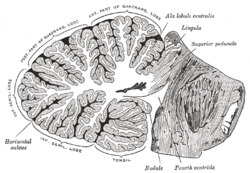
The cerebellar vermis is located in the medial, cortico-nuclear zone of the cerebellum, which is in the posterior fossa of the cranium. The primary fissure in the vermis curves ventrolaterally to the superior surface of the cerebellum, dividing it into anterior and posterior lobes. Functionally, the vermis is associated with bodily posture and locomotion. The vermis is included within the spinocerebellum and receives somatic sensory input from the head and proximal body parts via ascending spinal pathways.

The posterior cranial fossa is part of the cranial cavity, located between the foramen magnum and tentorium cerebelli. It contains the brainstem and cerebellum.

The medial rectus muscle is a muscle in the orbit.

The tympanic cavity is a small cavity surrounding the bones of the middle ear. Within it sit the ossicles, three small bones that transmit vibrations used in the detection of sound.
Horizontal fissure can refer to:

The lateral wall and the floor of the orbit are separated posteriorly by the inferior orbital fissure which transmits the zygomatic branch of the maxillary nerve and the ascending branches from the pterygopalatine ganglion. The infraorbital vessels are found in the inferior orbital fissure, and travel down the infraorbital groove into the infraorbital canal and exit through the infraorbital foramen. Inferior division of ophthalmic vein passes through the inferior orbital fissure.

The back part of the medial surface of the labyrinth of ethmoid is subdivided by a narrow oblique fissure, the superior meatus of the nose, bounded above by a thin, curved plate, the superior nasal concha.

Not to be confused with the inferior orbital fissure, which is just lateral to the infraorbital groove.

The porta hepatis or transverse fissure of the liver is a short but deep fissure, about 5 cm long, extending transversely beneath the left portion of the right lobe of the liver, nearer its posterior surface than its anterior border.

The tela choroidea is a region of meningeal pia mater and underlying ependyma that gives rise to the choroid plexus in each of the brain’s four ventricles. Tela is Latin for woven and is used to describe a web-like membrane or layer. The tela choroidea is a very thin part of the loose connective tissue of pia mater that overlies and closely adheres to the ependyma with no intervening tissue. It has a rich blood supply. The ependyma and vascular pia mater that make up the tela choroidea form regions of minute projections known as a choroid plexus that projects into each ventricle. The choroid plexus produces the cerebrospinal fluid of the ventricular system. The tela choroidea in the ventricles forms from different parts of the roof plate in the development of the embryo.
Transverse fissure can refer to:

The biventer lobule is a region of the cerebellum. It is triangular in shape; its apex points backward, and is joined by the gray band to the pyramid.
The folium vermis is a short, narrow, concealed band at the posterior extremity of the vermis, consisting apparently of a single folium, but in reality marked on its upper and under surfaces by secondary fissures.
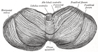
The central lobule is a small square lobule, situated in the anterior cerebellar notch. It overlaps the lingula, from which it is separated by the precentral fissure; laterally, it extends along the upper and anterior part of each hemisphere, where it forms a wing-like prolongation (ala), on each side, as the alae of the central lobule or alae lobuli centralis.

The monticulus of the cerebellum is divided by the primary fissure into an anterior, raised part, the culmen or summit, and a posterior sloped part, the clivus; the quadrangular lobule is similarly divided.
The culmen is the portion of the anterior vermis adjacent to the primary fissure of cerebellum.
The anatomy of the cerebellum can be viewed at three levels. At the level of gross anatomy, the cerebellum consists of a tightly folded and crumpled layer of cortex, with white matter underneath, several deep nuclei embedded in the white matter, and a fluid-filled ventricle in the middle. At the intermediate level, the cerebellum and its auxiliary structures can be broken down into several hundred or thousand independently functioning modules or "microzones". At the microscopic level, each module consists of the same small set of neuronal elements, laid out with a highly stereotyped geometry.