Basket cell

Last updated
Basket cell
Gray706.png
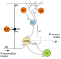
Transverse section of a cerebellar folium (Basket cell labeled at bottom left)
Details
Location Cerebellum
Shapemultipolar
FunctionInhibitory interneuron
Neurotransmitter GABA
Presynaptic connections Parallel fibers
Postsynaptic connections Purkinje cells
Identifiers
NeuroLex ID nifext_160
Anatomical terms of neuroanatomy

Basket cells are inhibitory GABAergic interneurons of the brain, found throughout different regions of the cortex and cerebellum. [1]

Contents

Anatomy and physiology

Basket cells are multipolar GABAergic interneurons that function to make inhibitory synapses and control the overall potentials of target cells. In general, dendrites of basket cells are free branching, contain smooth spines, and extend from 3 to 9 mm. Axons are highly branched, ranging in total from 20 to 50mm in total length. The branched axonal arborizations give rise to the name as they appear as baskets surrounding the soma of the target cell. [2] Basket cells form axo-somatic synapses, meaning their synapses target somas of other cells. [3] By controlling the somas of other neurons, basket cells can directly control the action potential discharge rate of target cells. [4]

Basket cells can be found throughout the brain, in among other the cortex, hippocampus, amygdala, basal ganglia, and the cerebellum.[ citation needed ]

Cortex

In the cortex, basket cells have sparsely branched axons giving off small pericellular, basket-shaped elaborations at several intervals along their length. Basket cells make up 5-10% of total neurons in the cortex. [5] There are three types of basket cells in the cortex, the small, large and nest type: [6] The axon of a small basket cell arborizes in the vicinity of that same cell's dendritic range, this axon is short. In contrast, large basket cells innervate somata in different cortical columns due to a long axon. [5] The nest basket cells are an intermediate form of the small and large cells, their axons are confined mainly to the same cortical layer as their somata. Nest basket cells have "radiating axonal collaterals" between that of large and small basket cells. They are included as basket cells because they are interneurons that perform axo-somatic synapses. [5]

Hippocampus

Hippocampal basket cells target somata and proximal dendrites of pyramidal neurons. Similar to their counterparts in the cortex, [7] hippocampal basket cells are also parvalbumin-expressing and fast-spiking. In the CA3 region of the hippocampus, basket cells can often form recurrent inhibition loops with pyramidal cells. [8] Projections from a pyramidal cell will innervate the basket cell, which in turn has a projection back onto the original pyramidal cells. Since basket cells are inhibitory, this generates a closed loop that can help dampen excitatory responses.

Cerebellum

Micrograph of cerebellar cortex showing Purkinje cells within the baskets formed by the processes of basket cells. Bielschowsky stain. Cerebellum - biel - very high mag.jpg
Micrograph of cerebellar cortex showing Purkinje cells within the baskets formed by the processes of basket cells. Bielschowsky stain.

In the cerebellum, the multipolar basket cells have branching dendrites, which are dilated and knotty. Basket cells synapse on the cell bodies of Purkinje cells and make inhibitory synapses with Purkinje cells. Cerebellar basket cell axons fire inhibitory neurotransmitters such as GABA to Purkinje cell axons, and inhibits the Purkinje cell. [9] Purkinje cells send inhibitory messages to the deep cerebellar nuclei and are responsible for the sole output of motor coordination from the cerebellar cortex. With the work of the basket cell, Purkinje cells do not send the inhibitory response for motor coordination and motor movement occurs. [10]

Additional images

See also

List of distinct cell types in the adult human body

Related Research Articles

<span class="mw-page-title-main">Neuron</span> Electrically excitable cell found in the nervous system of animals

Within a nervous system, a neuron, neurone, or nerve cell is an electrically excitable cell that fires electric signals called action potentials across a neural network. Neurons communicate with other cells via synapses - specialized connections that commonly use minute amounts of chemical neurotransmitters to pass the electric signal from the presynaptic neuron to the target cell through the synaptic gap. The neuron is the main component of nervous tissue in all animals except sponges and placozoa. Non-animals like plants and fungi do not have nerve cells.

<span class="mw-page-title-main">Cerebellum</span> Structure at the rear of the vertebrate brain, beneath the cerebrum

The cerebellum is a major feature of the hindbrain of all vertebrates. Although usually smaller than the cerebrum, in some animals such as the mormyrid fishes it may be as large as it or even larger. In humans, the cerebellum plays an important role in motor control. It may also be involved in some cognitive functions such as attention and language as well as emotional control such as regulating fear and pleasure responses, but its movement-related functions are the most solidly established. The human cerebellum does not initiate movement, but contributes to coordination, precision, and accurate timing: it receives input from sensory systems of the spinal cord and from other parts of the brain, and integrates these inputs to fine-tune motor activity. Cerebellar damage produces disorders in fine movement, equilibrium, posture, and motor learning in humans.

An inhibitory postsynaptic potential (IPSP) is a kind of synaptic potential that makes a postsynaptic neuron less likely to generate an action potential. IPSPs were first investigated in motorneurons by David P. C. Lloyd, John Eccles and Rodolfo Llinás in the 1950s and 1960s. The opposite of an inhibitory postsynaptic potential is an excitatory postsynaptic potential (EPSP), which is a synaptic potential that makes a postsynaptic neuron more likely to generate an action potential. IPSPs can take place at all chemical synapses, which use the secretion of neurotransmitters to create cell to cell signalling. Inhibitory presynaptic neurons release neurotransmitters that then bind to the postsynaptic receptors; this induces a change in the permeability of the postsynaptic neuronal membrane to particular ions. An electric current that changes the postsynaptic membrane potential to create a more negative postsynaptic potential is generated, i.e. the postsynaptic membrane potential becomes more negative than the resting membrane potential, and this is called hyperpolarisation. To generate an action potential, the postsynaptic membrane must depolarize—the membrane potential must reach a voltage threshold more positive than the resting membrane potential. Therefore, hyperpolarisation of the postsynaptic membrane makes it less likely for depolarisation to sufficiently occur to generate an action potential in the postsynaptic neurone.

<span class="mw-page-title-main">Purkinje cell</span> Specialized neuron in the cerebellum

Purkinje cells, or Purkinje neurons, are a class of GABAergic inhibitory neurons located in the cerebellum. They are named after their discoverer, Czech anatomist Jan Evangelista Purkyně, who characterized the cells in 1839.

An apical dendrite is a dendrite that emerges from the apex of a pyramidal cell. Apical dendrites are one of two primary categories of dendrites, and they distinguish the pyramidal cells from spiny stellate cells in the cortices. Pyramidal cells are found in the prefrontal cortex, the hippocampus, the entorhinal cortex, the olfactory cortex, and other areas. Dendrite arbors formed by apical dendrites are the means by which synaptic inputs into a cell are integrated. The apical dendrites in these regions contribute significantly to memory, learning, and sensory associations by modulating the excitatory and inhibitory signals received by the pyramidal cells.

<span class="mw-page-title-main">Dentate nucleus</span> Nucleus in the centre of each cerebellar hemisphere

The dentate nucleus is a cluster of neurons, or nerve cells, in the central nervous system that has a dentate – tooth-like or serrated – edge. It is located within the deep white matter of each cerebellar hemisphere, and it is the largest single structure linking the cerebellum to the rest of the brain. It is the largest and most lateral, or farthest from the midline, of the four pairs of deep cerebellar nuclei, the others being the globose and emboliform nuclei, which together are referred to as the interposed nucleus, and the fastigial nucleus. The dentate nucleus is responsible for the planning, initiation and control of voluntary movements. The dorsal region of the dentate nucleus contains output channels involved in motor function, which is the movement of skeletal muscle, while the ventral region contains output channels involved in nonmotor function, such as conscious thought and visuospatial function.

<span class="mw-page-title-main">Stellate cell</span>

Stellate cells are neurons in the central nervous system, named for their star-like shape formed by dendritic processes radiating from the cell body. Many stellate cells are GABAergic and are located in the molecular layer of the cerebellum. Stellate cells are derived from dividing progenitor cells in the white matter of postnatal cerebellum. Dendritic trees can vary between neurons. There are two types of dendritic trees in the cerebral cortex, which include pyramidal cells, which are pyramid shaped and stellate cells which are star shaped. Dendrites can also aid neuron classification. Dendrites with spines are classified as spiny, those without spines are classified as aspinous. Stellate cells can be spiny or aspinous, while pyramidal cells are always spiny. Most common stellate cells are the inhibitory interneurons found within the upper half of the molecular layer in the cerebellum. Cerebellar stellate cells synapse onto the dendritic trees of Purkinje cells and send inhibitory signals. Stellate neurons are sometimes found in other locations in the central nervous system; cortical spiny stellate cells are found in layer IVC of the primary visual cortex. In the somatosensory barrel cortex of mice and rats, glutamatergic (excitatory) spiny stellate cells are organized in the barrels of layer 4. They receive excitatory synaptic fibres from the thalamus and process feed forward excitation to 2/3 layer of the primary visual cortex to pyramidal cells. Cortical spiny stellate cells have a 'regular' firing pattern. Stellate cells are chromophobes, that is cells that does not stain readily, and thus appears relatively pale under the microscope.

<span class="mw-page-title-main">Golgi cell</span>

In neuroscience, Golgi cells are inhibitory interneurons found within the granular layer of the cerebellum. They were first identified as inhibitory in 1964. It was also the first example of an inhibitory feedback network, where the inhibitory interneuron was identified anatomically. These cells synapse onto the dendrite of granule cells and unipolar brush cells. They receive excitatory input from mossy fibres, also synapsing on granule cells, and parallel fibers, which are long granule cell axons. Thereby this circuitry allows for feed-forward and feed-back inhibition of granule cells.

Depolarization-induced suppression of inhibition is the classical and original electrophysiological example of endocannabinoid function in the central nervous system. Prior to the demonstration that depolarization-induced suppression of inhibition was dependent on the cannabinoid CB1 receptor function, there was no way of producing an in vitro endocannabinoid mediated effect.

The stratum lucidum of the hippocampus is a layer of the hippocampus between the stratum pyramidale and the stratum radiatum. It is the tract of the mossy fiber projections, both inhibitory and excitatory from the granule cells of the dentate gyrus. One mossy fiber may make up to 37 connections to a single pyramidal cell, and innervate around 12 pyramidal cells on top of that. Any given pyramidal cell in the stratum lucidum may get input from as many as 50 granule cells.

<span class="mw-page-title-main">Mossy fiber (hippocampus)</span> Pathway in the hippocampus

In the hippocampus, the mossy fiber pathway consists of unmyelinated axons projecting from granule cells in the dentate gyrus that terminate on modulatory hilar mossy cells and in Cornu Ammonis area 3 (CA3), a region involved in encoding short-term memory. These axons were first described as mossy fibers by Santiago Ramón y Cajal as they displayed varicosities along their lengths that gave them a mossy appearance. The axons that make up the pathway emerge from the basal portions of the granule cells and pass through the hilus of the dentate gyrus before entering the stratum lucidum of CA3. Granule cell synapses tend to be glutamatergic, though immunohistological data has indicated that some synapses contain neuropeptidergic elements including opiate peptides such as dynorphin and enkephalin. There is also evidence for co-localization of both GABAergic and glutamatergic neurotransmitters within mossy fiber terminals. GABAergic and glutamatergic co-localization in mossy fiber boutons has been observed primarily in the developing hippocampus, but in adulthood, evidence suggests that mossy fiber synapses may alternate which neurotransmitter is released through activity-dependent regulation.

<span class="mw-page-title-main">Chandelier cell</span>

Chandelier neurons or chandelier cells are a subset of GABAergic cortical interneurons. They are described as parvalbumin-containing and fast-spiking to distinguish them from other subtypes of GABAergic neurons, although more recent work has suggested that only a subset of chandelier cells test positive for parvalbumin by immunostaining. The name comes from the specific shape of their axon arbors, with the terminals forming distinct arrays called "cartridges". The cartridges are immunoreactive to an isoform of the GABA membrane transporter, GAT-1, and this serves as their identifying feature. GAT-1 is involved in the process of GABA reuptake into nerve terminals, thus helping to terminate its synaptic activity. Chandelier neurons synapse exclusively to the axon initial segment of pyramidal neurons, near the site where action potential is generated. It is believed that they provide inhibitory input to the pyramidal neurons, but there is data showing that in some circumstances the GABA from chandelier neurons could be excitatory.

<span class="mw-page-title-main">Hippocampus anatomy</span>

Hippocampus anatomy describes the physical aspects and properties of the hippocampus, a neural structure in the medial temporal lobe of the brain. It has a distinctive, curved shape that has been likened to the sea-horse monster of Greek mythology and the ram's horns of Amun in Egyptian mythology. This general layout holds across the full range of mammalian species, from hedgehog to human, although the details vary. For example, in the rat, the two hippocampi look similar to a pair of bananas, joined at the stems. In primate brains, including humans, the portion of the hippocampus near the base of the temporal lobe is much broader than the part at the top. Due to the three-dimensional curvature of this structure, two-dimensional sections such as shown are commonly seen. Neuroimaging pictures can show a number of different shapes, depending on the angle and location of the cut.

<span class="mw-page-title-main">Anatomy of the cerebellum</span> Structures in the cerebellum, a part of the brain

The anatomy of the cerebellum can be viewed at three levels. At the level of gross anatomy, the cerebellum consists of a tightly folded and crumpled layer of cortex, with white matter underneath, several deep nuclei embedded in the white matter, and a fluid-filled ventricle in the middle. At the intermediate level, the cerebellum and its auxiliary structures can be broken down into several hundred or thousand independently functioning modules or compartments known as microzones. At the microscopic level, each module consists of the same small set of neuronal elements, laid out with a highly stereotyped geometry.

<span class="mw-page-title-main">Granule cell</span> Type of neuron with a very small cell body

The name granule cell has been used for a number of different types of neurons whose only common feature is that they all have very small cell bodies. Granule cells are found within the granular layer of the cerebellum, the dentate gyrus of the hippocampus, the superficial layer of the dorsal cochlear nucleus, the olfactory bulb, and the cerebral cortex.

An autapse is a chemical or electrical synapse from a neuron onto itself. It can also be described as a synapse formed by the axon of a neuron on its own dendrites, in vivo or in vitro.

Lugaro cells are primary sensory interneurons of the cerebellum, that have an inhibitory function. They are fusiform, having a spindle shape that tapers at each end. They were first described by Ernesto Lugaro in the early 20th century. Lugaro cells are found just beneath the layer of Purkinje cells between the molecular layer and the granular layer. They have thick principal dendrites coming from opposite poles of their bodies. These dendrites are very long and travel along the boundary between the Purkinje layer and the granular layer. They seem to contact from 5 to 15 Purkinje cells in a horizontal direction. They can also sense stimuli close to the Purkinje cells and their dendrites form a large receptive area that monitors the environment near to the Purkinje cells. Whilst their dendrites make contact with the Purkinje cells they also receive inputs from branches of the Purkinje axons by which they seem to have a sampling and integration role.

Cartwheel cells are neurons of the dorsal cochlear nucleus (DCN) where they greatly outnumber the other inhibitory interneurons of the DCN. Their somas lie on the superficial side of the pyramidal layer of the DCN, and their dendrites receive input from the parallel fibres of the granule cell layer. Their axons do not extend beyond the dorsal cochlear nucleus but synapse with other cartwheel cells and pyramidal cells within the DCN releasing GABA and glycine onto their targets.

An axo-axonic synapse is a type of synapse, formed by one neuron projecting its axon terminals onto another neuron's axon.

<span class="mw-page-title-main">Brain cell</span> Functional tissue of the brain

Brain cells make up the functional tissue of the brain. The rest of the brain tissue is structural or connective called the stroma which includes blood vessels. The two main types of cells in the brain are neurons, also known as nerve cells, and glial cells also known as neuroglia.

References

  1. Jones, Edward (1984). Cerebral Cortex: Volume 1: Cellular Components of the Cerebral Cortex. Springer. ISBN   978-0-306-41544-9.
  2. "Stahl's Essential Pharmacology".[ unreliable medical source? ]
  3. Jones, EG; Hendry, SHC (1984). "Basket cells". In Peters, A; Jones, EG (eds.). Cerebral cortex: cellular components of the cerebral cortex. New York: Plenum Press. pp. 309–34.
  4. Cobb, S. R.; Buhl, E. H.; Halasy, K.; Paulsen, O.; Somogyi, P. (1995). "Synchronization of neuronal activity in hippocampus by individual GABAergic interneurons". Nature. 378 (6552): 75–8. Bibcode:1995Natur.378...75C. doi:10.1038/378075a0. PMID   7477292.
  5. 1 2 3 Wang, Y.; Gupta, A; Toledo-Rodriguez, M; Wu, C. Z.; Markram, H (2002). "Anatomical, Physiological, Molecular and Circuit Properties of Nest Basket Cells in the Developing Somatosensory Cortex". Cerebral Cortex. 12 (4): 395–410. doi: 10.1093/cercor/12.4.395 . PMID   11884355.
  6. Fox, Kevin. Barrel Cortex. Cambridge University Press. pp. 55–6.
  7. Contreras, Diego (2004). "Electrophysiological classes of neocortical neurons". Neural Networks. 17 (5–6): 633–46. doi:10.1016/j.neunet.2004.04.003. PMID   15288889.
  8. Bryne, John. "Feedback/recurrent inhibition: Feedback inhibition in microcircuits". Neuroscience Online. University of Texas Health Center. Archived from the original on 2013-12-03. Retrieved 2013-07-27.
  9. Southan, A. P.; Robertson, B (1998). "Patch-clamp recordings from cerebellar basket cell bodies and their presynaptic terminals reveal an asymmetric distribution of voltage-gated potassium channels". The Journal of Neuroscience. 18 (3): 948–55. PMID   9437016.
  10. Tan, Y. P.; Llano, I. (1999). "Modulation by K+channels of action potential-evoked intracellular Ca2+concentration rises in rat cerebellar basket cell axons". The Journal of Physiology. 520: 65–78. doi:10.1111/j.1469-7793.1999.00065.x. PMC   2269558 . PMID   10517801.