The medulla oblongata or simply medulla is a long stem-like structure which makes up part of the brainstem. It is anterior and partially inferior to the cerebellum. It is a cone-shaped neuronal mass responsible for autonomic (involuntary) functions ranging from vomiting to sneezing. The medulla contains the cardiac, respiratory, vomiting and vasomotor centers and therefore deals with the autonomic functions of breathing, heart rate and blood pressure as well as the sleep wake cycle.
In anatomy, a fissure is a groove, natural division, deep furrow, elongated cleft, or tear in various parts of the body also generally called a sulcus, or in the brain a sulcus.

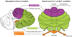
The cerebellar vermis is located in the medial, cortico-nuclear zone of the cerebellum, which is in the posterior fossa of the cranium. The primary fissure in the vermis curves ventrolaterally to the superior surface of the cerebellum, dividing it into anterior and posterior lobes. Functionally, the vermis is associated with bodily posture and locomotion. The vermis is included within the spinocerebellum and receives somatic sensory input from the head and proximal body parts via ascending spinal pathways.

The anterior lobe of cerebellum is the portion of the cerebellum responsible for mediating unconscious proprioception. Inputs into the anterior lobe of the cerebellum are mainly from the spinal cord.

The arbor vitae is the cerebellar white matter, so called for its branched, tree-like appearance. In some ways it more resembles a fern and is present in both cerebellar hemispheres. It brings sensory and motor information to and from the cerebellum. The arbor vitae is located deep in the cerebellum. Situated within the arbor vitae are the deep cerebellar nuclei; the dentate, globose, emboliform and the fastigial nuclei. These four different structures lead to the efferent projections of the cerebellum.

The dentate nucleus is a cluster of neurons, or nerve cells, in the central nervous system that has a dentate – tooth-like or serrated – edge. It is located within the deep white matter of each cerebellar hemisphere, and it is the largest single structure linking the cerebellum to the rest of the brain. It is the largest and most lateral, or farthest from the midline, of the four pairs of deep cerebellar nuclei, the others being the fastigial nucleus and the globose and emboliform nuclei which together are referred to as the interposed nucleus. The dentate nucleus is responsible for the planning, initiation and control of voluntary movements. The dorsal region of the dentate nucleus contains output channels involved in motor function, which is the movement of skeletal muscle, while the ventral region contains output channels involved in nonmotor function, such as conscious thought and visuospatial function.

The cerebellopontine angle (CPA) is located between the cerebellum and the pons. The cerebellopontine angle is the site of the cerebellopontine angle cistern one of the subarachnoid cisterns that contains cerebrospinal fluid, arachnoid tissue, cranial nerves, and associated vessels. The cerebellopontine angle is also the site of a set of neurological disorders known as the cerebellopontine angle syndrome.

Only a small part of the parieto-occipital sulcus, or parietooccipital fissure is seen on the lateral surface of the hemisphere, its chief part being on the medial surface.

The calcarine sulcus is an anatomical landmark located at the caudal end of the medial surface of the brain of humans and other primates. Its name comes from the Latin "calcar" meaning "spur". It is a complete sulcus.
The largest and deepest fissure in the cerebellum is named the horizontal fissure.
In the occipital bone, the lower division of the cruciate eminence is prominent, and is named the internal occipital crest; it bifurcates near the foramen magnum and gives attachment to the falx cerebelli; in the attached margin of this falx is the occipital sinus, which is sometimes duplicated.

Near the middle of the squamous part of occipital bone is the external occipital protuberance, the highest point of which is referred to as the inion. The inion is the most prominent projection of the protuberance which is located at the posterioinferior part of the human skull. The nuchal ligament and trapezius muscle attach to it.

In the human brain, the superior cerebellar peduncle is a paired structure of white matter that connects the cerebellum to the midbrain. It consists mainly of efferent fibers, the cerebellothalamic tract that runs from a cerebellar hemisphere to the contralateral thalamus, and the cerebellorubral tract that runs from a cerebellar hemisphere to the red nucleus. It also contains afferent tracts, most prominent of which is the ventral spinocerebellar tract. Other afferent tracts are the trigeminothalamic fibers, tectocerebellar fibers, and noradrenergic fibers from the locus coeruleus. The superior peduncle emerges from the upper and medial parts of the white matter of each hemisphere and is placed under cover of the upper part of the cerebellum.
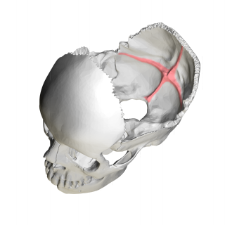
The cruciform eminence, divides the deeply concave internal surface of the occipital bone into four fossae:

The portion of the inferior frontal lobe immediately adjacent to the longitudinal fissure is named the straight gyrus,(or gyrus rectus) and is continuous with the superior frontal gyrus on the medial surface.

The biventer lobule is a region of the cerebellum. It is triangular in shape; its apex points backward, and is joined by the gray band to the pyramid.
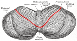
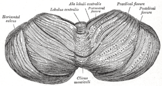
The central lobule is a small square lobule, situated in the anterior cerebellar notch. It overlaps the lingula, from which it is separated by the precentral fissure; laterally, it extends along the upper and anterior part of each hemisphere, where it forms a wing-like prolongation (ala), on each side, as the alae of the central lobule or alae lobuli centralis.

The posterior lobe of cerebellum or neocerebellum, is the portion of the cerebellum below the primary fissure.
The culmen is the portion of the anterior vermis adjacent to the primary fissure of cerebellum.

The anatomy of the cerebellum can be viewed at three levels. At the level of gross anatomy, the cerebellum consists of a tightly folded and crumpled layer of cortex, with white matter underneath, several deep nuclei embedded in the white matter, and a fluid-filled ventricle in the middle. At the intermediate level, the cerebellum and its auxiliary structures can be broken down into several hundred or thousand independently functioning modules or "microzones". At the microscopic level, each module consists of the same small set of neuronal elements, laid out with a highly stereotyped geometry.