Central lobule

Last updated
Central lobule
Gray703.png
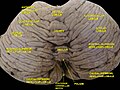
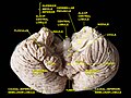
Anterior view of the cerebellum.
Details
Identifiers
Latin lobulus centralis
NeuroNames 658
NeuroLex ID birnlex_920
TA A14.1.07.105
FMA 72519
Anatomical terms of neuroanatomy

The central lobule is a small square lobule, situated in the anterior cerebellar notch. It overlaps the lingula, from which it is separated by the precentral fissure; laterally, it extends along the upper and anterior part of each hemisphere, where it forms a wing-like prolongation (ala), on each side, as the alae of the central lobule or alae lobuli centralis.

Lingula of cerebellum

The lingula is a small tongue-shaped process, consisting of four or five folia; it lies in front of the lobulus centralis, and is concealed by it.

Contents

Additional Images

Related Research Articles

Cerebellar vermis Anatomical structure in the brain

The cerebellar vermis is located in the medial, cortico-nuclear zone of the cerebellum, which resides in the posterior fossa of the cranium. The primary fissure in the vermis curves ventrolaterally to the superior surface of the cerebellum, dividing it into anterior and posterior lobes. Functionally, the vermis is associated with bodily posture and locomotion. The vermis is included within the spinocerebellum and receives somatic sensory input from the head and proximal body parts via ascending spinal pathways.

The vertebral arteries are major arteries of the neck. Typically, the vertebral arteries originate from the subclavian arteries. Each vessel courses superiorly along each side of the neck, merging within the skull to form the single, midline basilar artery. As the supplying component of the vertebrobasilar vascular system, the vertebral arteries provide supply blood to the upper spinal cord, brainstem, cerebellum, and posterior part of brain.

Calcarine sulcus

The calcarine sulcus is an anatomical landmark located at the caudal end of the medial surface of the brain. Its name comes from the Latin "calcar" meaning "spur". It is a complete sulcus.

Vestibular nuclei cranial nuclei for the vestibular nerve

The vestibular nuclei (VN) are the cranial nuclei for the vestibular nerve.

Facial colliculus

The facial colliculus is an elevated area located on the pontine tegmentum in the floor of the fourth ventricle. It is formed by fibers from the facial motor nucleus of the facial nerve as they loop over the abducens nucleus. Thus a lesion to the facial colliculus would result in ipsilateral facial paralysis and ipsilateral unopposed eye medial deviation.

Superior cerebellar peduncle

In the human brain, the superior cerebellar peduncle is a paired structure of white matter that connects the cerebellum to the midbrain. It consists mainly of efferent fibers, the cerebellothalamic tract that runs from a cerebellar hemisphere to the contralateral thalamus, and the cerebellorubral tract that runs from a cerebellar hemisphere to the red nucleus. It also contains afferent tracts, most prominent of which is the ventral spinocerebellar tract. Other afferent tracts are the trigeminothalamic fibers, tectocerebellar fibers, and noradrenergic fibers from the locus coeruleus. The superior peduncle emerges from the upper and medial parts of the white matter of each hemisphere and is placed under cover of the upper part of the cerebellum.

Middle cerebellar peduncle

The middle cerebellar peduncles are paired structures that connect the cerebellum to the pons and are composed entirely of centripetal fibers, i.e. incoming fibers. The fibers arise from the pontine nucleus to the opposite hemisphere of the cerebellar cortex. The fibers are arranged in three fasciculi: superior, inferior, and deep.

Lobe (anatomy) clear anatomical division or extension of an organ

In anatomy, a lobe is a clear anatomical division or extension of an organ that can be determined without the use of a microscope at the gross anatomy level. This is in contrast to the much smaller lobule, which is a clear division only visible under the microscope.

Vagal trigone

The cells of the dorsal nucleus of vagus nerve are spindle-shaped, like those of the posterior column of the spinal cord, and the nucleus is usually considered as representing the base of the posterior column. It measures about 2 cm. in length, and in the lower, closed part of the medulla oblongata is situated behind the dorsal motor nucleus of the vagus; whereas in the upper, open part it lies lateral to that nucleus, and corresponds to an eminence, named the vagal trigone, in the rhomboid fossa.

Cerebellar tonsil

The cerebellar tonsil is analogous to a rounded lobule on the undersurface of each cerebellar hemisphere, continuous medially with the uvula of the cerebellar vermis and superiorly by the flocculonodular lobe. Synonyms include: tonsilla cerebelli, amygdala cerebelli, the latter of which is not to be confused with the cerebral tonsils or amygdala nuclei located deep within the medial temporal lobes of the cerebral cortex. The flocculonodular lobe of the cerebellum which can also be confused for the cerebellar tonsils, is one of three lobes that make up the overall composition of the cerebellum. The cerebellum consists of three anatomical and functional lobes: anterior lobe, posterior lobe, and flocculonodular lobe.

Anterolateral sulcus of medulla

The anterolateral sulcus is a sulcus on the side of the medulla oblongata. The rootlets of cranial nerve XII emerge from this sulcus.

Hypoglossal trigone

In the upper part of the medulla oblongata, the hypoglossal nucleus approaches the rhomboid fossa, where it lies close to the middle line, under an eminence named the hypoglossal trigone. It is a slight elevation in the floor of the inferior recess of the fourth ventricle, beneath which is the nucleus of origin of the twelfth cranial nerve.

Nodule of vermis

The nodule, or anterior end of the inferior vermis, abuts against the roof of the fourth ventricle, and can only be distinctly seen after the cerebellum has been separated from the medulla oblongata and pons.

Tuber of vermis

The tuber of vermis, the most posterior division of the inferior vermis, is of small size, and laterally spreads out into the large inferior semilunar lobules, which comprise at least two-thirds of the inferior surface of the hemisphere.

Biventer lobule

The biventer lobule is a region of the cerebellum. It is triangular in shape; its apex points backward, and is joined by the gray band to the pyramid.

Primary fissure of cerebellum

The monticulus of the cerebellum is divided by the primary fissure into an anterior, raised part, the culmen or summit, and a posterior sloped part, the clivus; the quadrangular lobule is similarly divided.

Culmen (cerebellum) structure in the brain

The culmen is the portion of the anterior vermis adjacent to the primary fissure of cerebellum.

Uvula of cerebellum

The uvula forms a considerable portion of the inferior vermis; it is separated on either side from the tonsil by the sulcus vallecula, at the bottom of which it is connected to the tonsil by a ridge of gray matter, indented on its surface by shallow furrows, and hence called the furrowed band.

Orbital gyri

The inferior or orbital surface of the frontal lobe is concave, and rests on the orbital plate of the frontal bone. It is divided into four orbital gyri by a well-marked H-shaped orbital sulcus. These are named, from their position, the medial, anterior, lateral, and posterior, orbital gyri. The medial orbital gyrus presents a well-marked antero-posterior sulcus, the olfactory sulcus, for the olfactory tract; the portion medial to this is named the straight gyrus, and is continuous with the superior frontal gyrus on the medial surface.

References

This article incorporates text in the public domain from page 789 of the 20th edition of Gray's Anatomy (1918)

The public domain consists of all the creative works to which no exclusive intellectual property rights apply. Those rights may have expired, been forfeited, expressly waived, or may be inapplicable.

<i>Grays Anatomy</i> English-language textbook of human anatomy

Gray's Anatomy is an English language textbook of human anatomy originally written by Henry Gray and illustrated by Henry Vandyke Carter. Earlier editions were called Anatomy: Descriptive and Surgical and Gray's Anatomy: Descriptive and Applied, but the book's name is commonly shortened to, and later editions are titled, Gray's Anatomy. The book is widely regarded as an extremely influential work on the subject, and has continued to be revised and republished from its initial publication in 1858 to the present day. The latest edition of the book, the 41st, was published in September 2015.

Neuroscience Information Framework

The Neuroscience Information Framework is a repository of global neuroscience web resources, including experimental, clinical, and translational neuroscience databases, knowledge bases, atlases, and genetic/genomic resources and provides many authoritative links throughout the neuroscience portal of Wikipedia.