Related Research Articles

The cerebellum is a major feature of the hindbrain of all vertebrates. Although usually smaller than the cerebrum, in some animals such as the mormyrid fishes it may be as large as it or even larger. In humans, the cerebellum plays an important role in motor control and cognitive functions such as attention and language as well as emotional control such as regulating fear and pleasure responses, but its movement-related functions are the most solidly established. The human cerebellum does not initiate movement, but contributes to coordination, precision, and accurate timing: it receives input from sensory systems of the spinal cord and from other parts of the brain, and integrates these inputs to fine-tune motor activity. Cerebellar damage produces disorders in fine movement, equilibrium, posture, and motor learning in humans.

The medulla oblongata or simply medulla is a long stem-like structure which makes up the lower part of the brainstem. It is anterior and partially inferior to the cerebellum. It is a cone-shaped neuronal mass responsible for autonomic (involuntary) functions, ranging from vomiting to sneezing. The medulla contains the cardiac, respiratory, vomiting and vasomotor centers, and therefore deals with the autonomic functions of breathing, heart rate and blood pressure as well as the sleep–wake cycle. "Medulla" is from Latin, ‘pith or marrow’. And "oblongata" is from Latin, ‘lengthened or longish or elongated'.

The pons is part of the brainstem that in humans and other mammals, lies inferior to the midbrain, superior to the medulla oblongata and anterior to the cerebellum.
The brainstem is the posterior stalk-like part of the brain that connects the cerebrum with the spinal cord. In the human brain the brainstem is composed of the midbrain, the pons, and the medulla oblongata. The midbrain is continuous with the thalamus of the diencephalon through the tentorial notch, and sometimes the diencephalon is included in the brainstem.

In neuroanatomy, the trigeminal nerve (lit. triplet nerve), also known as the fifth cranial nerve, cranial nerve V, or simply CN V, is a cranial nerve responsible for sensation in the face and motor functions such as biting and chewing; it is the most complex of the cranial nerves. Its name (trigeminal, from Latin tri- 'three' and -geminus 'twin') derives from each of the two nerves (one on each side of the pons) having three major branches: the ophthalmic nerve (V1), the maxillary nerve (V2), and the mandibular nerve (V3). The ophthalmic and maxillary nerves are purely sensory, whereas the mandibular nerve supplies motor as well as sensory (or "cutaneous") functions. Adding to the complexity of this nerve is that autonomic nerve fibers as well as special sensory fibers (taste) are contained within it.

In neuroanatomy, a neural pathway is the connection formed by axons that project from neurons to make synapses onto neurons in another location, to enable neurotransmission. Neurons are connected by a single axon, or by a bundle of axons known as a nerve tract, or fasciculus. Shorter neural pathways are found within grey matter in the brain, whereas longer projections, made up of myelinated axons, constitute white matter.

The dorsal column–medial lemniscus pathway (DCML) (also known as the posterior column-medial lemniscus pathway is the major sensory pathway of the central nervous system that conveys sensations of fine touch, vibration, two-point discrimination, and proprioception from the skin and joints. It transmits this information to the somatosensory cortex of the postcentral gyrus in the parietal lobe of the brain. The pathway receives information from sensory receptors throughout the body, and carries this in the gracile fasciculus and the cuneate fasciculus, tracts that make up the white matter dorsal columns of the spinal cord. At the level of the medulla oblongata, the fibers of the tracts decussate and are continued in the medial lemniscus, on to the thalamus and relayed from there through the internal capsule and transmitted to the somatosensory cortex. The name dorsal-column medial lemniscus comes from the two structures that carry the sensory information: the dorsal columns of the spinal cord, and the medial lemniscus in the brainstem.

The medial lemniscus, also known as Reil's band or Reil's ribbon, is a large ascending bundle of heavily myelinated axons that decussate in the brainstem, specifically in the medulla oblongata. The medial lemniscus is formed by the crossings of the internal arcuate fibers. The internal arcuate fibers are composed of axons of the gracile nucleus and the cuneate nucleus. The cell bodies of the nuclei lie contralaterally.

The inferior olivary nucleus (ION) is a structure found in the medulla oblongata underneath the superior olivary nucleus. In vertebrates, the ION is known to coordinate signals from the spinal cord to the cerebellum to regulate motor coordination and learning. These connections have been shown to be tightly associated, as degeneration of either the cerebellum or the ION results in degeneration of the other.

The spinocerebellar tracts are nerve tracts originating in the spinal cord and terminating in the same side (ipsilateral) of the cerebellum. The two main tracts are the dorsal spinocerebellar tract, and the ventral spinocerebellar tract. Both of these tracts are located in the peripheral region of the lateral funiculi.

The inferior cerebellar peduncle is formed by fibers of the restiform body that join with fibers from the much smaller juxtarestiform body. The inferior cerebellar peduncle is the smallest of the three cerebellar peduncles.

A cranial nerve nucleus is a collection of neurons in the brain stem that is associated with one or more of the cranial nerves. Axons carrying information to and from the cranial nerves form a synapse first at these nuclei. Lesions occurring at these nuclei can lead to effects resembling those seen by the severing of nerve(s) they are associated with. All the nuclei except that of the trochlear nerve supply nerves of the same side of the body.

The vestibular nuclei (VN) are the cranial nuclei for the vestibular nerve located in the brainstem.

The lateral vestibular nucleus is the continuation upward and lateralward of the principal nucleus, and in it terminate many of the ascending branches of the vestibular nerve.
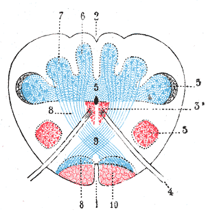
The dorsal column nuclei are a pair of nuclei in the dorsal columns of the dorsal column–medial lemniscus pathway (DCML) in the brainstem. The name refers collectively to the cuneate nucleus and gracile nucleus, which are situated at the lower end of the medulla oblongata. Both nuclei contain second-order neurons of the DCML, which convey fine touch and proprioceptive information from the body to the brain via the thalamus.
The ventral trigeminal tract, ventral trigeminothalamic tract, anterior trigeminal tract, or anterior trigeminothalamic tract, is a tract composed of second-order neuronal axons. These afferent fibers carry sensory information about discriminative and crude touch, conscious proprioception, pain, and temperature from the head, face, and oral cavity. The ventral trigeminal tract connects the two major components of the brainstem trigeminal complex – the principal, or main sensory nucleus and the spinal trigeminal nucleus, to the ventral posteromedial nucleus of the thalamus.

The trigeminal lemniscus or the trigeminothalamic tracts is a somatosensory tract containing second-order neuron fibers of the trigeminal system. It consists of the ventral and dorsal trigeminal tracts. Its second-order sensory axons convey tactile, pain, and temperature impulses from the skin of the face, the mucous membranes of the nasal and oral cavities, and the eye, as well as proprioceptive information from the facial and masticatory muscles.
The dorsal trigeminal tract are uncrossed second-order sensory fibers conveying fine (discriminative) touch and pressure information from the dorsomedial division of principal sensory nucleus of trigeminal nerve to the ipsilateral ventral posteromedial nucleus of thalamus. Second-order fibers from the ventrolateral division of the principal sensory nucleus meanwhile cross-over to ascend contralaterally in the ventral trigeminal tract along with those fibers arising from the spinal trigeminal nucleus.
The anatomy of the cerebellum can be viewed at three levels. At the level of gross anatomy, the cerebellum consists of a tightly folded and crumpled layer of cortex, with white matter underneath, several deep nuclei embedded in the white matter, and a fluid-filled ventricle in the middle. At the intermediate level, the cerebellum and its auxiliary structures can be broken down into several hundred or thousand independently functioning modules or compartments known as microzones. At the microscopic level, each module consists of the same small set of neuronal elements, laid out with a highly stereotyped geometry.

The following diagram is provided as an overview of and topical guide to the human nervous system:
References
- ↑ Haines, Duane (2018). Fundamental neuroscience for basic and clinical applications (Fifth ed.). Philadelphia, PA: Elsevier. p. 395. ISBN 9780323396325.