Nodule of vermis

Last updated
Nodule of vermis
CerebellumDiv.png
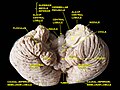
Schematic representation of the major anatomical subdivisions of the cerebellum. Superior view of an "unrolled" cerebellum, placing the vermis in one plane.
Gray704.png
Sagittal section of the cerebellum, near the junction of the vermis with the hemisphere. (Nodule labeled at bottom right.)
Details
Identifiers
Latin nodulus vermis
NeuroNames 681
TA A14.1.07.302
FMA 83882
Anatomical terms of neuroanatomy

The nodule (nodular lobe), or anterior end of the inferior vermis, abuts against the roof of the fourth ventricle, and can only be distinctly seen after the cerebellum has been separated from the medulla oblongata and pons.

Fourth ventricle

The fourth ventricle is one of the four connected fluid-filled cavities within the human brain. These cavities, known collectively as the ventricular system, consist of the left and right lateral ventricles, the third ventricle, and the fourth ventricle. The fourth ventricle extends from the cerebral aqueduct to the obex, and is filled with cerebrospinal fluid (CSF).

Cerebellum region of the brain that coordinates motor functions and muscle tone

The cerebellum is a major feature of the hindbrain of all vertebrates. Although usually smaller than the cerebrum, in some animals such as the mormyrid fishes it may be as large as or even larger. In humans, the cerebellum plays an important role in motor control. It may also be involved in some cognitive functions such as attention and language as well as in regulating fear and pleasure responses, but its movement-related functions are the most solidly established. The human cerebellum does not initiate movement, but contributes to coordination, precision, and accurate timing: it receives input from sensory systems of the spinal cord and from other parts of the brain, and integrates these inputs to fine-tune motor activity. Cerebellar damage produces disorders in fine movement, equilibrium, posture, and motor learning in humans.

Medulla oblongata structure of the brain stem

The medulla oblongata is a long stem-like structure located in the brainstem. It is anterior and partially inferior to the cerebellum. It is a cone-shaped neuronal mass responsible for autonomic (involuntary) functions ranging from vomiting to sneezing. The medulla contains the cardiac, respiratory, vomiting and vasomotor centers and therefore deals with the autonomic functions of breathing, heart rate and blood pressure.

On either side of the nodule is a thin layer of white substance, named the posterior medullary velum.

It is semilunar in form, its convex border being continuous with the white substance of the cerebellum; it extends on either side as far as the flocculus.

Flocculus (cerebellar)

The flocculus is a small lobe of the cerebellum at the posterior border of the middle cerebellar peduncle anterior to the biventer lobule. Like other parts of the cerebellum, the flocculus is involved in motor control. It is an essential part of the vestibulo-ocular reflex, and aids in the learning of basic motor skills in the brain.

Additional Images


Related Research Articles

Inferior cerebellar peduncle

The upper part of the posterior district of the medulla oblongata is occupied by the inferior cerebellar peduncle, a thick rope-like strand situated between the lower part of the fourth ventricle and the roots of the glossopharyngeal and vagus nerves.

Dentate nucleus

The dentate nucleus is a cluster of neurons, or nerve cells, in the central nervous system that has a dentate – tooth-like or serrated – edge. It is located within the deep white matter of each cerebellar hemisphere, and it is the largest single structure linking the cerebellum to the rest of the brain. It is the largest and most lateral, or farthest from the midline, of the four pairs of deep cerebellar nuclei, the others being the fastigial nucleus and the globose and emboliform nuclei which together are referred to as the interposed nucleus. The dentate nucleus is responsible for the planning, initiation and control of voluntary movements. The dorsal region of the dentate nucleus contains output channels involved in motor function, which is the movement of skeletal muscle, while the ventral region contains output channels involved in nonmotor function, such as conscious thought and visuospatial function.

Cerebellar tentorium A vertebrate brain structure separating the cerebellum from the occipital lobes

The cerebellar tentorium or tentorium cerebelli is an extension of the dura mater that separates the cerebellum from the inferior portion of the occipital lobes.

Vestibular nuclei cranial nuclei for the vestibular nerve

The vestibular nuclei (VN) are the cranial nuclei for the vestibular nerve.

Squamous part of occipital bone

The squamous part of occipital bone, is situated above and behind the foramen magnum, and is curved from above downward and from side to side.

Cerebellar peduncles connect the cerebellum to the brain stem. There are six cerebellar peduncles in total, three on each side:

Superior cerebellar peduncle

In the human brain, the superior cerebellar peduncle is a paired structure of white matter that connects the cerebellum to the midbrain. It consists mainly of efferent fibers, the cerebellothalamic tract that runs from a cerebellar hemisphere to the contralateral thalamus, and the cerebellorubral tract that runs from a cerebellar hemisphere to the red nucleus. It also contains afferent tracts, most prominent of which is the ventral spinocerebellar tract. Other afferent tracts are the trigeminothalamic fibers, tectocerebellar fibers, and noradrenergic fibers from the locus coeruleus. The superior peduncle emerges from the upper and medial parts of the white matter of each hemisphere and is placed under cover of the upper part of the cerebellum.

Middle cerebellar peduncle

The middle cerebellar peduncles are paired structures that connect the cerebellum to the pons and are composed entirely of centripetal fibers, i.e. incoming fibers. The fibers arise from the pontine nucleus to the opposite hemisphere of the cerebellar cortex. The fibers are arranged in three fasciculi: superior, inferior, and deep.

Superior medullary velum

The superior medullary velum is a thin, transparent lamina of white matter, which stretches between the superior cerebellar peduncles; on the dorsal surface of its lower half the folia and lingula are prolonged.

Inferior medullary velum

The inferior medullary velum is a thin layer of white substance, prolonged from the white center of the cerebellum, above and on either side of the nodule; it forms the infero-posterior part of the fourth ventricle.

Vallecula of cerebellum

On the superior surface of cerebellum, the vermis protrudes above the level of the hemispheres, but on the inferior surface it is sunk almost out of sight in the bottom of a deep depression between them; this depression is called the vallecula of the cerebellum, and lodges the posterior part of the medulla oblongata and the inferior vermis, which consists of the tuber vermis, pyramid, uvula and nodule.

Biventer lobule

The biventer lobule is a region of the cerebellum. It is triangular in shape; its apex points backward, and is joined by the gray band to the pyramid.

Central lobule

The central lobule is a small square lobule, situated in the anterior cerebellar notch. It overlaps the lingula, from which it is separated by the precentral fissure; laterally, it extends along the upper and anterior part of each hemisphere, where it forms a wing-like prolongation (ala), on each side, as the alae of the central lobule or alae lobuli centralis.

Pontocerebellar fibers

The pontocerebellar fibers are the second order neuron fibers of the corticopontocerebellar tracts that cross to the other side of the pons and run within the middle cerebellar peduncles, from the pons to the contralateral cerebellum.

Culmen (cerebellum) structure in the brain

The culmen is the portion of the anterior vermis adjacent to the primary fissure of cerebellum.

Lingula of cerebellum

The lingula is a small tongue-shaped process, consisting of four or five folia; it lies in front of the lobulus centralis, and is concealed by it.

Uvula of cerebellum

The uvula forms a considerable portion of the inferior vermis; it is separated on either side from the tonsil by the sulcus vallecula, at the bottom of which it is connected to the tonsil by a ridge of gray matter, indented on its surface by shallow furrows, and hence called the furrowed band.