Superior cerebellar peduncle

Last updated
Superior cerebellar peduncle
Gray704.png
Sagittal section of the cerebellum, near the junction of the vermis with the hemisphere. (Superior peduncle labeled at upper right.)
Gray705.png
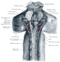
Dissection showing the projection fibers of the cerebellum. (Superior peduncle labeled at center top.)
Details
Identifiers
Latin pedunculus cerebellaris superior
NeuroNames 833
NeuroLex ID birnlex_1711
TA98 A14.1.05.006
A14.1.07.417
A14.1.08.678
A14.1.06.009
A14.1.06.216
TA2 5846
FMA 72495
Anatomical terms of neuroanatomy

In the human brain, the superior cerebellar peduncle (brachium conjunctivum) is one of the three paired cerebellar peduncles of bundled fibers that connect the cerebellum to the brainstem. The superior cerebellar peduncle connects to the midbrain. It consists mainly of efferent fibers, the cerebellothalamic tract that runs from a cerebellar hemisphere to the contralateral thalamus, and the cerebellorubral tract that runs from a cerebellar hemisphere to the red nucleus. It also contains afferent tracts, most prominent of which is the ventral spinocerebellar tract. Other afferent tracts are the ventral trigeminal tract, tectocerebellar fibers, and noradrenergic fibers from the locus coeruleus. The superior peduncle emerges from the upper and medial parts of the white matter of each cerebellar hemisphere[ citation needed ] and is placed under cover of the upper part of the cerebellum.

Contents

Structure

Superior cerebellar peduncles are connected together by the anterior medullary velum, which can be followed upward as far as the inferior colliculi, under which they disappear.

Below, they form the upper lateral boundaries of the fourth ventricle, but as they ascend they converge on the dorsal aspect of the ventricle and thus assist in forming its roof.

Decussation

Deep dissection of brain-stem showing decussation Gray690.png
Deep dissection of brain-stem showing decussation

The decussation of superior cerebellar peduncle is the crossing of fibers of the superior cerebellar peduncle across the midline, and is located at the level of the inferior colliculi. It comprises the cerebellothalamic tract, which arises from the dentate nucleus (therefore also known as dentatothalamic tract), as well as the cerebellorubral tract, which arises from the globose and emboliform nuclei and project to the contralateral red nucleus to eventually become the rubrospinal tract. It is also known as horseshoe-shaped commissure of Wernekinck . [1] It is important as an anatomical landmark, as lesions above it cause contralateral cerebellar signs, while lesions below it cause ipsilateral cerebellar signs.

Additional images

Related Research Articles

Articles related to anatomy include:

<span class="mw-page-title-main">Pons</span> Part of the brainstem in humans and other bipeds

The pons is part of the brainstem that in humans and other mammals, lies inferior to the midbrain, superior to the medulla oblongata and anterior to the cerebellum.

<span class="mw-page-title-main">Brainstem</span> Posterior part of the brain, adjoining and structurally continuous

The brainstem is the posterior stalk-like part of the brain that connects the cerebrum with the spinal cord. In the human brain the brainstem is composed of the midbrain, the pons, and the medulla oblongata. The midbrain is continuous with the thalamus of the diencephalon through the tentorial notch, and sometimes the diencephalon is included in the brainstem.

<span class="mw-page-title-main">Midbrain</span> Forward-most portion of the brainstem

The midbrain or mesencephalon is the rostral-most portion of the brainstem connecting the diencephalon and cerebrum with the pons. It consists of the cerebral peduncles, tegmentum, and tectum.

<span class="mw-page-title-main">Inferior colliculus</span> Midbrain structure involved in the auditory pathway

The inferior colliculus (IC) is the principal midbrain nucleus of the auditory pathway and receives input from several peripheral brainstem nuclei in the auditory pathway, as well as inputs from the auditory cortex. The inferior colliculus has three subdivisions: the central nucleus, a dorsal cortex by which it is surrounded, and an external cortex which is located laterally. Its bimodal neurons are implicated in auditory-somatosensory interaction, receiving projections from somatosensory nuclei. This multisensory integration may underlie a filtering of self-effected sounds from vocalization, chewing, or respiration activities.

<span class="mw-page-title-main">Inferior olivary nucleus</span> Brain structure in the medulla that helps coordinate movement

The inferior olivary nucleus (ION) is a structure found in the medulla oblongata underneath the superior olivary nucleus. In vertebrates, the ION is known to coordinate signals from the spinal cord to the cerebellum to regulate motor coordination and learning. These connections have been shown to be tightly associated, as degeneration of either the cerebellum or the ION results in degeneration of the other.

<span class="mw-page-title-main">Spinocerebellar tracts</span> Nerve tract in humans

The spinocerebellar tracts are nerve tracts originating in the spinal cord and terminating in the same side (ipsilateral) of the cerebellum. The two main tracts are the dorsal spinocerebellar tract, and the ventral spinocerebellar tract. Both of these tracts are located in the peripheral region of the lateral funiculi.

<span class="mw-page-title-main">Inferior cerebellar peduncle</span>

The inferior cerebellar peduncle is formed by fibers of the restiform body that join with fibers from the much smaller juxtarestiform body. The inferior cerebellar peduncle is the smallest of the three cerebellar peduncles.

The interposed nucleus is the combined globose and emboliform nuclei on either side. The interposed nucleus is one of the paired cerebellar nuclei. It is located in the roof of the fourth ventricle, lateral to the fastigial nucleus. The emboliform nucleus is the anterior interposed nucleus, and the globose nucleus is the posterior interposed nucleus.

<span class="mw-page-title-main">Posterior cerebral artery</span> Artery which supplies blood to the occipital lobe of the brain

The posterior cerebral artery (PCA) is one of a pair of cerebral arteries that supply oxygenated blood to the occipital lobe, part of the back of the human brain. The two arteries originate from the distal end of the basilar artery, where it bifurcates into the left and right posterior cerebral arteries. These anastomose with the middle cerebral arteries and internal carotid arteries via the posterior communicating arteries.

<span class="mw-page-title-main">Cerebellar peduncles</span> Structure connecting the cerebellum to the brainstem

The cerebellar peduncles are three paired bundles of fibres that connect the cerebellum to the brain stem.

<span class="mw-page-title-main">Middle cerebellar peduncle</span> Structure in the brain connecting the pons to the cerebellum

The middle cerebellar peduncle is one of three paired cerebellar peduncles connecting the brainstem to the cerebellum. The connection is from the pons. It connects the pons to the cerebellum, with fibres originating from the pontine nuclei, and travelling to the opposite cerebellar hemisphere. It is supplied by the anterior inferior cerebellar artery (AICA) and branches from the basilar artery. It conveys information from the cerebrum and the pons to the cerebellum.

<span class="mw-page-title-main">Myoclonic triangle</span>

The myoclonic triangle is an important feedback circuit of the brainstem and deep cerebellar nuclei which is responsible for modulating spinal cord motor activity.

<span class="mw-page-title-main">Pontocerebellar fibers</span>

The pontocerebellar fibers are the second-order neuron fibers of the corticopontocerebellar tracts that cross to the other side of the pons and run within the middle cerebellar peduncles, from the pons to the contralateral cerebellum. They arise from the pontine nuclei as the second part of the corticopontocerebellar tract, and decussate (cross-over) in the pons before passing through the middle cerebellar peduncles to reach and terminate in the contralateral posterior lobe of the cerebellum (neocerebellum). It is part of a pathway involved in the coordination of voluntary movements.

The cerebellothalamic tract or the tractus cerebellothalamicus, is part of the superior cerebellar peduncle. It originates in the cerebellar nuclei, crosses completely in the decussation of the superior cerebellar peduncle, bypasses the red nucleus, and terminates in posterior division of ventral lateral nucleus of thalamus. The ventrolateral nucleus has different divisions and distinct connections, mostly with frontal and parietal lobes. The primary motor cortex and premotor cortex get information from the ventrolateral nucleus projections originating in the interposed nucleus and dentate nuclei. Other dentate nucleus projections via thalamic pathway transmit information to prefrontal cortex and posterior parietal cortex. The cerebellum sends thalamocortical projections and in addition may also send connections from the thalamus to association areas serving cognitive and affective functions.

<span class="mw-page-title-main">Anatomy of the cerebellum</span> Structures in the cerebellum, a part of the brain

The anatomy of the cerebellum can be viewed at three levels. At the level of gross anatomy, the cerebellum consists of a tightly folded and crumpled layer of cortex, with white matter underneath, several deep nuclei embedded in the white matter, and a fluid-filled ventricle in the middle. At the intermediate level, the cerebellum and its auxiliary structures can be broken down into several hundred or thousand independently functioning modules or compartments known as microzones. At the microscopic level, each module consists of the same small set of neuronal elements, laid out with a highly stereotyped geometry.

<span class="mw-page-title-main">Anatomical terms of neuroanatomy</span> Terminology used to describe the central and peripheral nervous systems

This article describes anatomical terminology that is used to describe the central and peripheral nervous systems - including the brain, brainstem, spinal cord, and nerves.

Friedrich Christian Gregor Wernekinck was a German anatomist. His specialties were anatomy and mineralogy. Wernekinck was born in Münster in 1798. After attending the University of Münster and University of Göttingen, he received his doctorate at the University of Giessen, where he became a full professor of philosophy in 1826. He died in 1835.

The nucleus of Darkschewitsch is an accessory oculomotor nucleus situated in the ventrolateral portion of the periaqueductal gray of the mesencephalon (midbrain) near its junction with the diencephalon. It is involved in mediating vertical eye movements. It projects to the trochlear nucleus, receives afferents from the visual cortex, and forms a reciprocal (looping) connection with the cerebellum by way of the inferior olive.

References

PD-icon.svgThis article incorporates text in the public domain from page 792 of the 20th edition of Gray's Anatomy (1918)

  1. Voogd J, van Baarsen K (February 2014). "The horseshoe-shaped commissure of Wernekinck or the decussation of the brachium conjunctivum methodological changes in the 1840s". The Cerebellum. 13 (1): 113–20. doi:10.1007/s12311-013-0520-9. PMID   24078481. S2CID   15717777.