Midbrain tegmentum

Last updated
Midbrain tegmentum
Gray712.png
Transverse section of mid-brain at level of superior colliculi. ("Tegmentum" visible center right.)
Cn3nucleus.png
Section through superior colliculus showing path of oculomotor nerve. (Tegmentum not labeled, but surrounding structures more clearly defined.)
Details
Part of Midbrain
Identifiers
Latin Tegmentum Mesencephali
MeSH D013681
NeuroNames 491
NeuroLex ID birnlex_1200
Anatomical terms of neuroanatomy

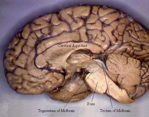
The midbrain is anatomically delineated into the tectum (roof) and the tegmentum (floor). The midbrain tegmentum extends from the substantia nigra to the cerebral aqueduct in a horizontal section of the midbrain. It forms the floor of the midbrain that surrounds below the cerebral aqueduct as well as the floor of the fourth ventricle while the midbrain tectum forms the roof of the fourth ventricle. The tegmentum contains a collection of tracts and nuclei with movement-related, species-specific, and pain-perception functions. The general structures of midbrain tegmentum include red nucleus and the periaqueductal grey matter.

Contents

Nigrostriatal Pathway and Mesolimbic Pathway in the Dopaminergic System Nigrostriatal pathway.svg
Nigrostriatal Pathway and Mesolimbic Pathway in the Dopaminergic System

Unlike the midbrain tectum (which is a sensory structure located posteriorly), the midbrain tegmentum, which locates anteriorly, is related to a number of motor functions. Within the tegmentum, the red nucleus is in charge of motor coordination (specifically for limb movements) and the periaqueductal gray matter (PAG) contains critical circuits for modulating behavioral responses to pains. The substantia nigra (black substance) serves an important role in rewarding behaviors such as approaching desired objects. In addition, the substantia nigra forms reciprocal connections with the basal ganglia which are highly correlated with motor functions and learning.

The midbrain tegmentum is also an important part of the dopaminergic system which is essential for feelings of reward and pleasure. Two regions in the midbrain tegmentum are of particular interest. The first one is the substantia nigra which is an important part of the nigrostriatal pathway. This pathway serves to coordinate motor movements and when left unbalanced, motor deficits would follow. For instance, when the dopamine neurons are lost from the substantia nigra, the condition of extreme muscle rigidity occurs as in the Parkinson's disease. The second region is the ventral tegmental area (VTA; or simply ventral tegmentum) which is at the hub of the mesolimbic pathway. Specifically, the VTA is the origin of dopaminergic cell bodies from which signals reach the anterior parts of the brain (e.g., frontal lobes) as well as the posterior parts (e.g., cerebellum). Because of this pathway regulates the experience of reward and pleasure, it is not surprising to see that food and drugs affect it the most in terms of a loss of impulse control. That is, the mesolimbic pathway is essential in regulating drug addiction. The potential mechanism is through the associative learning of the environmental cues and reward. For instance, through each drug use, individuals increasingly associate the cues related to each drug use (e.g., the room in which the drug is taken or the people with which individuals take drug). Over time, the drug enhances the dopamine-related, classically-conditioned cues associated with drug taking. As a result, later encounters with these cues will produce and heighten dopamine activity and subsequently prompt individuals to crave drugs. Moreover, excessive mesolimbic dopamine activity plays a role in schizophrenia, a behavioral disorder characterized by delusions, hallucinations, blunted emotion, agitation, etc. On the other hand, a lack of mesolimbic dopamine activity may induce deficits in attention. [1]

See also

Notes

  1. Kolb, Bryan; Ian Q. Whishaw. (2015). Fundamentals of Human Neuropsychology (7. ed.). New York, NY: Macmillan. p. 71-G-30. ISBN   978-1-4292-8295-6.

Related Research Articles

Striatum Nucleus in the basal ganglia of the brain

The striatum, or corpus striatum, is a nucleus in the subcortical basal ganglia of the forebrain. The striatum is a critical component of the motor and reward systems; receives glutamatergic and dopaminergic inputs from different sources; and serves as the primary input to the rest of the basal ganglia.

Substantia nigra Structure in the basal ganglia of the brain

The substantia nigra (SN) is a basal ganglia structure located in the midbrain that plays an important role in reward and movement. Substantia nigra is Latin for "black substance", reflecting the fact that parts of the substantia nigra appear darker than neighboring areas due to high levels of neuromelanin in dopaminergic neurons. Parkinson's disease is characterized by the loss of dopaminergic neurons in the substantia nigra pars compacta.

Dopamine Organic chemical that functions both as a hormone and a neurotransmitter

Dopamine is a neuromodulatory molecule that plays several important roles in cells. It is an organic chemical of the catecholamine and phenethylamine families. Dopamine constitutes about 80% of the catecholamine content in the brain. It is an amine synthesized by removing a carboxyl group from a molecule of its precursor chemical, L-DOPA, which is synthesized in the brain and kidneys. Dopamine is also synthesized in plants and most animals. In the brain, dopamine functions as a neurotransmitter—a chemical released by neurons to send signals to other nerve cells. Neurotransmitters are synthesized in specific regions of the brain, but affect many regions systemically. The brain includes several distinct dopamine pathways, one of which plays a major role in the motivational component of reward-motivated behavior. The anticipation of most types of rewards increases the level of dopamine in the brain, and many addictive drugs increase dopamine release or block its reuptake into neurons following release. Other brain dopamine pathways are involved in motor control and in controlling the release of various hormones. These pathways and cell groups form a dopamine system which is neuromodulatory.

Basal ganglia Group of subcortical nuclei involved in the motor and reward systems

The basal ganglia are a group of subcortical nuclei, of varied origin, in the brains of vertebrates. In humans, and some primates, there are some differences, mainly in the division of the globus pallidus into an external and internal region, and in the division of the striatum. The basal ganglia are situated at the base of the forebrain and top of the midbrain. Basal ganglia are strongly interconnected with the cerebral cortex, thalamus, and brainstem, as well as several other brain areas. The basal ganglia are associated with a variety of functions, including control of voluntary motor movements, procedural learning, habit learning, conditional learning, eye movements, cognition, and emotion.

Brainstem Posterior part of the brain, adjoining and structurally continuous

The brainstem is the posterior stalk-like part of the brain that connects the cerebrum with the spinal cord. In the human brain the brainstem is composed of the midbrain, the pons, and the medulla oblongata. The midbrain is continuous with the thalamus of the diencephalon through the tentorial notch, and sometimes the diencephalon is included in the brainstem.

The mesolimbic pathway, sometimes referred to as the reward pathway, is a dopaminergic pathway in the brain. The pathway connects the ventral tegmental area in the midbrain to the ventral striatum of the basal ganglia in the forebrain. The ventral striatum includes the nucleus accumbens and the olfactory tubercle.

Midbrain Forward-most portion of the brainstem

The midbrain or mesencephalon is the forward-most portion of the brainstem and is associated with vision, hearing, motor control, sleep and wakefulness, arousal (alertness), and temperature regulation. The name comes from the Greek mesos, "middle", and enkephalos, "brain".

Nucleus accumbens Region of the basal forebrain

The nucleus accumbens is a region in the basal forebrain rostral to the preoptic area of the hypothalamus. The nucleus accumbens and the olfactory tubercle collectively form the ventral striatum. The ventral striatum and dorsal striatum collectively form the striatum, which is the main component of the basal ganglia. The dopaminergic neurons of the mesolimbic pathway project onto the GABAergic medium spiny neurons of the nucleus accumbens and olfactory tubercle. Each cerebral hemisphere has its own nucleus accumbens, which can be divided into two structures: the nucleus accumbens core and the nucleus accumbens shell. These substructures have different morphology and functions.

Dopaminergic pathways Projection neurons in the brain that synthesize and release dopamine

Dopaminergic pathways, in the human brain are involved in both physiological and behavioral processes including movement, cognition, executive functions, reward, motivation, and neuroendocrine control. Each pathway is a set of projection neurons, consisting of individual dopamine neurons.

Oculomotor nucleus

The fibers of the oculomotor nerve arise from a nucleus in the midbrain, which lies in the gray substance of the floor of the cerebral aqueduct and extends in front of the aqueduct for a short distance into the floor of the third ventricle. From this nucleus the fibers pass forward through the tegmentum, the red nucleus, and the medial part of the substantia nigra, forming a series of curves with a lateral convexity, and emerge from the oculomotor sulcus on the medial side of the cerebral peduncle.

Nigrostriatal pathway

The nigrostriatal pathway is a bilateral dopaminergic pathway in the brain that connects the substantia nigra pars compacta (SNc) in the midbrain with the dorsal striatum in the forebrain. It is one of the four major dopamine pathways in the brain, and is critical in the production of movement as part of a system called the basal ganglia motor loop. Dopaminergic neurons of this pathway release dopamine from axon terminals that synapse onto GABAergic medium spiny neurons (MSNs), also known as spiny projection neurons (SPNs), located in the striatum.

Mesocortical pathway

The mesocortical pathway is a dopaminergic pathway that connects the ventral tegmentum to the prefrontal cortex. It is one of the four major dopamine pathways in the brain. It is essential to the normal cognitive function of the dorsolateral prefrontal cortex, and is thought to be involved in cognitive control, motivation, and emotional response.

Ventral tegmental area Group of neurons on the floor of the midbrain

The ventral tegmental area (VTA), also known as the ventral tegmental area of Tsai, or simply ventral tegmentum, is a group of neurons located close to the midline on the floor of the midbrain. The VTA is the origin of the dopaminergic cell bodies of the mesocorticolimbic dopamine system and other dopamine pathways; it is widely implicated in the drug and natural reward circuitry of the brain. The VTA plays an important role in a number of processes, including reward cognition and orgasm, among others, as well as several psychiatric disorders. Neurons in the VTA project to numerous areas of the brain, ranging from the prefrontal cortex to the caudal brainstem and several regions in between.

Tegmentum

The tegmentum is a general area within the brainstem. The tegmentum is the ventral part of the midbrain and the tectum is the dorsal part of the midbrain. It is located between the ventricular system and distinctive basal or ventral structures at each level. It forms the floor of the midbrain (mesencephalon) whereas the tectum forms the ceiling. It is a multisynaptic network of neurons that is involved in many subconscious homeostatic and reflexive pathways. It is a motor center that relays inhibitory signals to the thalamus and basal nuclei preventing unwanted body movement.

The pars compacta is a portion of the substantia nigra, located in the midbrain. It is formed by dopaminergic neurons and located medial to pars reticulata. Parkinson's disease is characterized by the death of dopaminergic neurons in this region.

Reward system Group of neural structures responsible for motivation and desire

The reward system is a group of neural structures responsible for incentive salience, associative learning, and positively-valenced emotions, particularly ones involving pleasure as a core component. Reward is the attractive and motivational property of a stimulus that induces appetitive behavior, also known as approach behavior, and consummatory behavior. A rewarding stimulus has been described as "any stimulus, object, event, activity, or situation that has the potential to make us approach and consume it is by definition a reward". In operant conditioning, rewarding stimuli function as positive reinforcers; however, the converse statement also holds true: positive reinforcers are rewarding.

The ventral pallidum (VP) is a structure within the basal ganglia of the brain. It is an output nucleus whose fibres project to thalamic nuclei, such as the ventral anterior nucleus, the ventral lateral nucleus, and the medial dorsal nucleus. The VP is a core component of the reward system which forms part of the limbic loop of the basal ganglia, a pathway involved in the regulation of motivational salience, behavior, and emotions. It is involved in addiction.

Dopaminergic cell groups are collections of neurons in the central nervous system that synthesize the neurotransmitter dopamine. In the 1960s, dopamine neurons were first identified and named by Annica Dahlström and Kjell Fuxe, who used histochemical fluorescence. The subsequent discovery of genes encoding enzymes that synthesize dopamine, and transporters that incorporate dopamine into synaptic vesicles or reclaim it after synaptic release, enabled scientists to identify dopaminergic neurons by labeling gene or protein expression that is specific to these neurons.

The rostromedial tegmental nucleus (RMTg), also known as the tail of the ventral tegmental area (tVTA), is a GABAergic nucleus which functions as a "master brake" for the midbrain dopamine system. It is poorly differentiated from the rest of the ventral tegmental area (VTA) and possesses robust functional and structural links to the dopamine pathways. Notably, both acute and chronic exposure to psychostimulants have been shown to induce FosB and ΔFosB expression in the RMTg; no other drug type has been shown to induce these proteins in the RMTg.