You can help expand this article with text translated from the corresponding article in Portuguese. (November 2025)Click [show] for important translation instructions.
|
| Cerebral peduncle | |
|---|---|
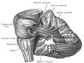
 Superficial dissection of brain-stem. Ventral view. ("cerebral peduncle" visible in red at center-right) | |
 Obtuse section (perpendicular to the brainstem) through superior colliculus showing path of oculomotor nerve (crus cerebri labeled at lower left). | |
| Details | |
| Identifiers | |
| Latin | pedunculus cerebri |
| MeSH | D065850 |
| NeuroNames | 487 |
| NeuroLex ID | birnlex_1202 |
| TA98 | A14.1.06.004 |
| TA2 | 5878 |
| FMA | 62394 |
| Anatomical terms of neuroanatomy | |
The cerebral peduncles (In Latin, ped- means 'foot'.) are the two stalks that attach the cerebrum to the brainstem. [1] They are structures at the front of the midbrain which arise from the ventral pons and contain the large ascending (sensory) and descending (motor) tracts that run to and from the cerebrum from the pons. Mainly, the three common areas that give rise to the cerebral peduncles are the cerebral cortex, the spinal cord and the cerebellum. [2] The region includes the tegmentum, crus cerebri and pretectum. By this definition, the cerebral peduncles are also known as the basis pedunculi, while the large ventral bundle of efferent fibers is referred to as the cerebral crus (crus means ‘leg’ in Latin.) or the pes pedunculi (pes means 'foot' in Latin.).
Contents
The cerebral peduncles are located on either side of the midbrain and are the frontmost part of the midbrain, and act as the connectors between the rest of the midbrain and the thalamic nuclei and thus the cerebrum. As a whole, the cerebral peduncles assist in refining motor movements, learning new motor skills, and converting proprioceptive information into balance and posture maintenance. [3] [4] Important fiber tracts that run through the cerebral peduncles are the corticospinal, corticopontine, and corticobulbar tracts. Damage to the cerebral peduncles results in unrefined motor skills, imbalance, and lack of proprioception.[ medical citation needed ]


