
The levator ani is a broad, thin muscle group, situated on either side of the pelvis. It is formed from three muscle components: the pubococcygeus, the iliococcygeus, and the puborectalis.

Connective tissue is one of the four primary types of animal tissue, along with epithelial tissue, muscle tissue, and nervous tissue. It develops mostly from the mesenchyme, derived from the mesoderm, the middle embryonic germ layer. Connective tissue is found in between other tissues everywhere in the body, including the nervous system. The three meninges, membranes that envelop the brain and spinal cord, are composed of connective tissue. Most types of connective tissue consists of three main components: elastic and collagen fibers, ground substance, and cells. Blood, and lymph are classed as specialized fluid connective tissues that do not contain fiber. All are immersed in the body water. The cells of connective tissue include fibroblasts, adipocytes, macrophages, mast cells and leukocytes.

The Purkinje fibers, named for Jan Evangelista Purkyně, are located in the inner ventricular walls of the heart, just beneath the endocardium in a space called the subendocardium. The Purkinje fibers are specialized conducting fibers composed of electrically excitable cells. They are larger than cardiomyocytes with fewer myofibrils and many mitochondria. They conduct cardiac action potentials more quickly and efficiently than any of the other cells in the heart's electrical conduction system. Purkinje fibers allow the heart's conduction system to create synchronized contractions of its ventricles, and are essential for maintaining a consistent heart rhythm.
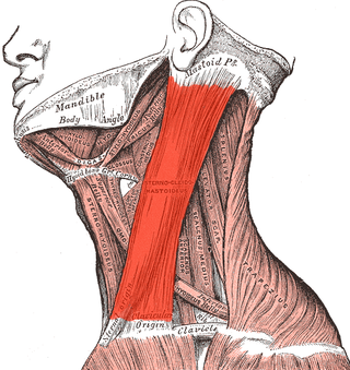
The sternocleidomastoid muscle is one of the largest and most superficial cervical muscles. The primary actions of the muscle are rotation of the head to the opposite side and flexion of the neck. The sternocleidomastoid is innervated by the accessory nerve.

The deltoid muscle is the muscle forming the rounded contour of the human shoulder. It is also known as the 'common shoulder muscle', particularly in other animals such as the domestic cat. Anatomically, the deltoid muscle is made up of three distinct sets of muscle fibers, namely the
- anterior or clavicular part
- posterior or scapular part
- intermediate or acromial part

The ciliary muscle is an intrinsic muscle of the eye formed as a ring of smooth muscle in the eye's middle layer, uvea. It controls accommodation for viewing objects at varying distances and regulates the flow of aqueous humor into Schlemm's canal. It also changes the shape of the lens within the eye but not the size of the pupil which is carried out by the sphincter pupillae muscle and dilator pupillae.

The levator labii superioris is a muscle of the human body used in facial expression. It is a broad sheet, the origin of which extends from the side of the nose to the zygomatic bone.

General visceral efferent fibers (GVE), visceral efferents or autonomic efferents are the efferent nerve fibers of the autonomic nervous system that provide motor innervation to smooth muscle, cardiac muscle, and glands through postganglionic varicosities.

The superior hypogastric plexus is a plexus of nerves situated on the vertebral bodies anterior to the bifurcation of the abdominal aorta. It bifurcates to form the left and the right hypogastric nerve. The SHP is the continuation of the abdominal aortic plexus.

The short ciliary nerves are nerves of the orbit around the eye. They are branches of the ciliary ganglion. They supply parasympathetic and sympathetic nerve fibers to the ciliary muscle, iris, and cornea. Damage to the short ciliary nerve may result in loss of the pupillary light reflex, or mydriasis.
Thoracic splanchnic nerves are splanchnic nerves that arise from the sympathetic trunk in the thorax and travel inferiorly to provide sympathetic supply to the abdomen. The nerves contain preganglionic sympathetic fibers and general visceral afferent fibers.

The frontopontine fibers or frontopontine tract are corticopontine fibers projecting from the cortex of the frontal lobe to the pons. In the internal capsule, the fibers descend predominately in the anterior limb, passing inferior to the thalamus to reach the mesencephalon (midbrain) where they descend in the medial portion of base of the cerebral peduncles. In the pons, the fibers flare out between the pontine nuclei.
Pelvic splanchnic nerves or nervi erigentes are splanchnic nerves that arise from sacral spinal nerves S2, S3, S4 to provide parasympathetic innervation to the organs of the pelvic cavity.

The esophageal plexus is formed by nerve fibers from two sources, branches of the vagus nerve, and visceral branches of the sympathetic trunk. The esophageal plexus and the cardiac plexus contain the same types of fibers and are both considered thoracic autonomic plexus.

The hypogastric nerves are the continuation of the superior hypogastric plexus that descend into the pelvis anterior the sacrum and become the inferior hypogastric plexuses on either side of pelvic organs. The hypogastric nerves serve as a pathway for autonomic fibers to communicate between the lower abdomen and pelvis.
The general visceral afferent (GVA) fibers conduct sensory impulses from the internal organs, glands, and blood vessels to the central nervous system. They are considered to be part of the visceral nervous system, which is closely related to the autonomic nervous system, but 'visceral nervous system' and 'autonomic nervous system' are not direct synonyms and care should be taken when using these terms. Unlike the efferent fibers of the autonomic nervous system, the afferent fibers are not classified as either sympathetic or parasympathetic.
Special visceral afferent fibers (SVA) are afferent fibers that develop in association with the gastrointestinal tract. They carry the special sense of taste (gustation). The cranial nerves containing SVA fibers are the facial nerve (VII), the glossopharyngeal nerve (IX), and the vagus nerve (X). The facial nerve receives taste from the anterior 2/3 of the tongue; the glossopharyngeal from the posterior 1/3, and the vagus nerve from the epiglottis. The sensory processes, using their primary cell bodies from the inferior ganglion, send projections to the medulla, from which they travel in the tractus solitarius, later terminating at the rostral nucleus solitarius.
Special somatic afferent fibers (SSA) are the afferent nerve fibers that carry information from the special senses of vision, hearing and balance. The cranial nerves containing SSA fibers are the optic nerve and the vestibulocochlear nerve.

A nerve tract is a bundle of nerve fibers (axons) connecting nuclei of the central nervous system. In the peripheral nervous system, this is known as a nerve fascicle, and has associated connective tissue. The main nerve tracts in the central nervous system are of three types: association fibers, commissural fibers, and projection fibers. A nerve tract may also be referred to as a commissure, decussation, or neural pathway. A commissure connects the two cerebral hemispheres at the same levels, while a decussation connects at different levels.
Beta motor neurons, also called beta motoneurons, are a few kind of lower motor neuron, along with alpha motor neurons and gamma motor neurons. Beta motor neurons innervate intrafusal fibers of muscle spindles with collaterals to extrafusal fibers - a type of slow twitch fiber. Also, axons of alpha, beta, and gamma motor neurons become myelinated. Moreover, these efferent neurons originate from the anterior grey column of the spinal cord and travel to skeletal muscles. However, the larger diameter alpha motor fibers require higher conduction velocity than beta and gamma.
