The brainstem is the stalk-like part of the brain that interconnects the cerebrum and diencephalon with the spinal cord. In the human brain, the brainstem is composed of the midbrain, the pons, and the medulla oblongata. The midbrain is continuous with the thalamus of the diencephalon through the tentorial notch.

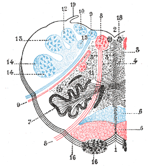
The hypoglossal nerve, also known as the twelfth cranial nerve, cranial nerve XII, or simply CN XII, is a cranial nerve that innervates all the extrinsic and intrinsic muscles of the tongue except for the palatoglossus, which is innervated by the vagus nerve. CN XII is a nerve with a sole motor function. The nerve arises from the hypoglossal nucleus in the medulla as a number of small rootlets, pass through the hypoglossal canal and down through the neck, and eventually passes up again over the tongue muscles it supplies into the tongue.

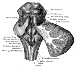
The hypoglossal nucleus is a cranial nerve nucleus, found within the medulla. Being a motor nucleus, it is close to the midline. In the open medulla, it is visible as what is known as the hypoglossal trigone, a raised area protruding slightly into the fourth ventricle.

Medial medullary syndrome, also known as inferior alternating syndrome, hypoglossal alternating hemiplegia, lower alternating hemiplegia, or Dejerine syndrome, is a type of alternating hemiplegia characterized by a set of clinical features resulting from occlusion of the anterior spinal artery. This results in the infarction of medial part of the medulla oblongata.

The fourth ventricle is one of the four connected fluid-filled cavities within the human brain. These cavities, known collectively as the ventricular system, consist of the left and right lateral ventricles, the third ventricle, and the fourth ventricle. The fourth ventricle extends from the cerebral aqueduct to the obex, and is filled with cerebrospinal fluid (CSF).

In anatomy, the olivary bodies or simply olives are a pair of prominent oval structures in the medulla oblongata, the lower portion of the brainstem. They contain the olivary nuclei.

The trigone is a smooth triangular region of the internal urinary bladder formed by the two ureteric orifices and the internal urethral meatus.

The fastigial nucleus is located in the cerebellum. It is one of the four deep cerebellar nuclei, and is grey matter embedded in the white matter of the cerebellum.

The ansa cervicalis is a loop formed by muscular branches of the cervical plexus formed by branches of cervical spinal nerves C1-C3. The ansa cervicalis has two roots - a superior root and an inferior root - that unite distally, forming a loop. It is situated within the carotid sheath.

The genioglossus is one of the paired extrinsic muscles of the tongue. It is a fan-shaped muscle that comprises the bulk of the body of the tongue. It arises from the mental spine of the mandible; it inserts onto the hyoid bone, and the bottom of the tongue. It is innervated by the hypoglossal nerve. The genioglossus is the major muscle responsible for protruding the tongue.

The hypoglossal canal is a foramen in the occipital bone of the skull. It is hidden medially and superiorly to each occipital condyle. It transmits the hypoglossal nerve.

The vestibular nuclei (VN) are the cranial nuclei for the vestibular nerve located in the brainstem.

The nucleus raphe obscurus, despite the implications of its name, has some very specific functions and connections of afferent and efferent nature. The nucleus raphes obscurus projects to the cerebellar lobes VI and VII and to crus II along with the nucleus raphe pontis.

The rhomboid fossa is a rhombus-shaped depression that is the anterior part of the fourth ventricle. Its anterior wall, formed by the back of the pons and the medulla oblongata, constitutes the floor of the fourth ventricle.

The vagal trigone is a triangular eminence upon the rhomboid fossa produced by the underlying dorsal nucleus of vagus nerve.

The emboliform nucleus is a deep cerebellar nucleus that lies immediately to the medial side of the nucleus dentatus, and partly covering its hilum. It is one among the four pairs of deep cerebellar nuclei, which are from lateral to medial: the dentate, interposed, and fastigial nuclei. These nuclei can be seen using Weigert's elastic stain.

The habenular commissure is a nerve tract of commissural fibers that connects the habenular nuclei on both sides of the habenular trigone in the epithalamus.
The habenular trigone is a small depressed triangular area situated anterior/superior to the superior colliculus. It contains the habenular nuclei.
James Thomas Wilson FRS (1861-1945) was a Professor of Anatomy at the University of Cambridge and an elected Fellow of the Royal Society.


