Septum pellucidum

Last updated
Septum pellucidum
Gray732.png
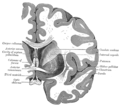
Image showing the septum pellucidum, with other structures of the rhinencephalon also shown
Gray720.png
Cross-section of the brain showing the right cerebral hemisphere. The septum pellucidum is seen as the sheet joining the corpus callosum to the fornix.
Details
LocationMidline of the brain
Identifiers
Latin Septum pellucidum
(lamina septi pellucidi)
MeSH D012688
NeuroNames 256
NeuroLex ID nlx_144186
TA98 A14.1.09.262
TA2 5647
FMA 61844
Anatomical terms of neuroanatomy

The septum pellucidum (Latin for "translucent wall") is a thin, triangular, vertical double membrane separating the anterior horns of the left and right lateral ventricles of the brain. It runs as a sheet from the corpus callosum down to the fornix.

Contents

The septum is not present in the syndrome septo-optic dysplasia.

Structure

The septum pellucidum is located in the septal area in the midline of the brain between the two cerebral hemispheres. The septal area is also the location of the septal nuclei. It is attached to the lower part of the corpus callosum, the large collection of nerve fibers that connect the two cerebral hemispheres. It is attached to the front forward part of the fornix. The lateral ventricles sit on either side of the septum.

The septum pellucidum consists of two layers or laminae of both white and gray matter. [1] During fetal development, there is a space between the two laminae called the cave of septum pellucidum that, in ninety percent of cases, disappears during infancy. [2] [3] The cavum was occasionally referred to as the fifth ventricle, but this is no longer used because the space is usually not continuous with the ventricular system. [4] The fifth ventricle is recognised as the terminal enlargement of the spinal cord. [5]

Clinical significance

Absence of the septum pellucidum occurs in septo-optic dysplasia, a rare developmental disorder usually characterized by abnormal development of the optic disk and pituitary deficiencies. [6] Symptoms of septo-optic dysplasia are highly variable and may include vision difficulties, low muscle tone, hormonal problems, seizures, intellectual problems, and jaundice at birth. [6] Management is directed at the symptoms a person is affected with. [6]

Additional images

See also

Related Research Articles

Articles related to anatomy include:

Colpocephaly Medical condition

Colpocephaly is a cephalic disorder involving the disproportionate enlargement of the occipital horns of the lateral ventricles and is usually diagnosed early after birth due to seizures. It is a nonspecific finding and is associated with multiple neurological syndromes, including agenesis of the corpus callosum, Chiari malformation, lissencephaly, and microcephaly. Although the exact cause of colpocephaly is not known yet, it is commonly believed to occur as a result of neuronal migration disorders during early brain development, intrauterine disturbances, perinatal injuries, and other central nervous system disorders. Individuals with colpocephaly have various degrees of motor disabilities, visual defects, spasticity, and moderate to severe intellectual disability. No specific treatment for colpocephaly exists, but patients may undergo certain treatments to improve their motor function or intellectual disability.

Septo-optic dysplasia Medical condition

Septo-optic dysplasia (SOD), known also as de Morsier syndrome, is a rare congenital malformation syndrome that features a combination of the underdevelopment of the optic nerve, pituitary gland dysfunction, and absence of the septum pellucidum . Two or more of these features need to be present for a clinical diagnosis — only 30% of patients have all three. French-Swiss doctor Georges de Morsier first recognized the relation of a rudimentary or absent septum pellucidum with hypoplasia of the optic nerves and chiasm in 1956.

Corpus callosum White matter tract connecting the two cerebral hemispheres

The corpus callosum, also callosal commissure, is a wide, thick nerve tract, consisting of a flat bundle of commissural fibers, beneath the cerebral cortex in the brain. The corpus callosum is only found in placental mammals. It spans part of the longitudinal fissure, connecting the left and right cerebral hemispheres, enabling communication between them. It is the largest white matter structure in the human brain, about ten centimetres in length and consisting of 200–300 million axonal projections.

Ventricular system

The ventricular system is a set of four interconnected cavities known as cerebral ventricles in the brain. Within each ventricle is a region of choroid plexus which produces the circulating cerebrospinal fluid (CSF). The ventricular system is continuous with the central canal of the spinal cord from the fourth ventricle, allowing for the flow of CSF to circulate.

Cerebral hemisphere The left and right cerebral hemispheres of the brain

The vertebrate cerebrum (brain) is formed by two cerebral hemispheres that are separated by a groove, the longitudinal fissure. The brain can thus be described as being divided into left and right cerebral hemispheres. Each of these hemispheres has an outer layer of grey matter, the cerebral cortex, that is supported by an inner layer of white matter. In eutherian (placental) mammals, the hemispheres are linked by the corpus callosum, a very large bundle of nerve fibers. Smaller commissures, including the anterior commissure, the posterior commissure and the fornix, also join the hemispheres and these are also present in other vertebrates. These commissures transfer information between the two hemispheres to coordinate localized functions.

Fornix (neuroanatomy)

The fornix is a C-shaped bundle of nerve fibers in the brain that acts as the major output tract of the hippocampus. The fornix also carries some afferent fibers to the hippocampus from structures in the diencephalon and basal forebrain. The fornix is part of the limbic system. While its exact function and importance in the physiology of the brain are still not entirely clear, it has been demonstrated in humans that surgical transection – the cutting of the fornix along its body – can cause memory loss. There is some debate over what type of memory is affected by this damage, but it has been found to most closely correlate with recall memory rather than recognition memory. This means that damage to the fornix can cause difficulty in recalling long-term information such as details of past events, but it has little effect on the ability to recognize objects or familiar situations.

Optic nerve hypoplasia (ONH) is a medical condition arising from the underdevelopment of the optic nerve(s). This condition is the most common congenital optic nerve anomaly. The optic disc appears abnormally small, because not all the optic nerve axons have developed properly. It is often associated with endocrinopathies, developmental delay, and brain malformations. The optic nerve, which is responsible for transmitting visual signals from the retina to the brain, has approximately 1.2 million nerve fibers in the average person. In those diagnosed with ONH, however, there are noticeably fewer nerves.

Longitudinal fissure

The longitudinal fissure is the deep groove that separates the two cerebral hemispheres of the vertebrate brain. Lying within it is a continuation of the dura mater called the falx cerebri. The inner surfaces of the two hemispheres are convoluted by gyri and sulci just as is the outer surface of the brain.

Lateral ventricles Two largest ventricles in each cerebral hemisphere

The lateral ventricles are the two largest ventricles of the brain and contain cerebrospinal fluid (CSF). Each cerebral hemisphere contains a lateral ventricle, known as the left or right ventricle, respectively.

Interventricular foramina (neuroanatomy) It is part of diencephalon that makes connection between lateral and third ventricular

In the brain, the interventricular foramina are channels that connect the paired lateral ventricles with the third ventricle at the midline of the brain. As channels, they allow cerebrospinal fluid (CSF) produced in the lateral ventricles to reach the third ventricle and then the rest of the brain's ventricular system. The walls of the interventricular foramina also contain choroid plexus, a specialized CSF-producing structure, that is continuous with that of the lateral and third ventricles above and below it.

Agenesis of the corpus callosum (ACC) is a rare birth defect in which there is a complete or partial absence of the corpus callosum. It occurs when the development of the corpus callosum, the band of white matter connecting the two hemispheres in the brain, in the embryo is disrupted. The result of this is that the fibers that would otherwise form the corpus callosum are instead longitudinally oriented along the ipsilateral ventricular wall and form structures called Probst bundles.

Posterior cerebral artery

The posterior cerebral artery (PCA) is one of a pair of arteries that supply oxygenated blood to the occipital lobe, part of the back of the human brain. The two arteries originate from the distal end of the basilar artery, where it bifurcates into the left and right posterior cerebral arteries. These anastomose with the middle cerebral arteries and internal carotid arteries via the posterior communicating arteries.

Dandy–Walker malformation Congenital malformation of the cerebellar vermis

Dandy–Walker malformation (DWM), also known as Dandy–Walker syndrome (DWS), is a rare congenital brain malformation in which the part joining the two hemispheres of the cerebellum does not fully form, and the fourth ventricle and space behind the cerebellum are enlarged with cerebrospinal fluid. Most of those affected develop hydrocephalus within the first year of life, which can present as increasing head size, vomiting, excessive sleepiness, irritability, downward deviation of the eyes and seizures. Other, less common symptoms are generally associated with comorbid genetic conditions and can include congenital heart defects, eye abnormalities, intellectual disability, congenital tumours, other brain defects such as agenesis of the corpus callosum, skeletal abnormalities, an occipital encephalocele or underdeveloped genitalia or kidneys. It is sometimes discovered in adolescents or adults due to mental health problems.

Septal area

The septal area is an area in the lower, posterior part of the medial surface of the frontal lobe, and refers to the nearby septum pellucidum.

Tela choroidea

The tela choroidea is a region of meningeal pia mater that adheres to the underlying ependyma, and gives rise to the choroid plexus in each of the brain’s four ventricles. Tela is Latin for woven and is used to describe a web-like membrane or layer. The tela choroidea is a very thin part of the loose connective tissue of pia mater overlying and closely adhering to the ependyma. It has a rich blood supply. The ependyma and vascular pia mater – the tela choroidea, form regions of minute projections known as a choroid plexus that projects into each ventricle. The choroid plexus produces most of the cerebrospinal fluid of the central nervous system that circulates through the ventricles of the brain, the central canal of the spinal cord, and the subarachnoid space. The tela choroidea in the ventricles forms from different parts of the roof plate in the development of the embryo.

Commissural fiber axons that connect the two hemispheres of the brain

The commissural fibers or transverse fibers are axons that connect the two hemispheres of the brain. In contrast to commissural fibers, association fibers connect regions within the same hemisphere of the brain, and projection fibers connect each region to other parts of the brain or to the spinal cord.

Cave of septum pellucidum

The cave of septum pellucidum (CSP), cavum septi pellucidi, or cavity of septum pellucidum is a slit-like space in the septum pellucidum that is present in fetuses but usually fuses during infancy. The septum pellucidum is a thin, laminated translucent vertical membrane in the midline of the brain separating the anterior horns of the right and left ventricles. It lies posterior to the corpus callosum. Persistence of the cave of septum pellucidum after infancy has been loosely associated with neural maldevelopment and several mental disorders that correlate with decreased brain tissue.

Septum verum

Septum Verum is a region in the lower medial part of the telencephalon that separates the two cerebral hemispheres. The human septum consists of two parts: the septum pellucidum, a thin membrane consisting of white matter and glial cells that separate the lateral ventricles, and the lower, precommisural septum verum, which consists of nuclei and grey matter. The term is sometimes used synonymously with Area Septalis, to refer to the precommisural part of the lower base of the telencephalon. The Septum verum contains the septal nuclei, which are usually considered part of the limbic system. 

References

  1. Lennart Heimer; Gary W. Van Hoesen (16 November 2007). Anatomy of neuropsychiatry: the new anatomy of the basal forebrain and its implications for neuropsychiatric illness. Academic Press. p. 28. ISBN   978-0-12-374239-1 . Retrieved 24 December 2010.
  2. David L. Clark; Nash N. Boutros; Mario F. Mendez (2010). The Brain and Behavior: An Introduction to Behavioral Neuroanatomy. Cambridge University Press. pp. 217–8. ISBN   978-0-521-14229-8 . Retrieved 24 December 2010.
  3. Love J; Hollenhorst R (1956). "Bilateral palsy of the sixth cranial nerve caused by a cyst of the septum pellucidum (fifth ventricle) and cured by pneumoencephalography". Mayo Clin Proc. 31 (2): 43–6. PMID   13289891.
  4. Alonso J; Coveñas R; Lara J; Piñuela C; Aijón J (1989). "The cavum septi pellucidi: a fifth ventricle?". Acta Anat (Basel). 134 (4): 286–90. doi:10.1159/000146704. PMID   2741657.
  5. Liccardo G; Ruggeri F; De Cerchio L; Floris R; Lunardi P (2005). "Fifth ventricle: an unusual cystic lesion of the conus medullaris". Spinal Cord. 43 (6): 381–4. doi: 10.1038/sj.sc.3101712 . PMID   15655569.
  6. 1 2 3 "Septo-optic dysplasia spectrum | Genetic and Rare Diseases Information Center (GARD) – an NCATS Program". rarediseases.info.nih.gov. Retrieved 2020-01-15.