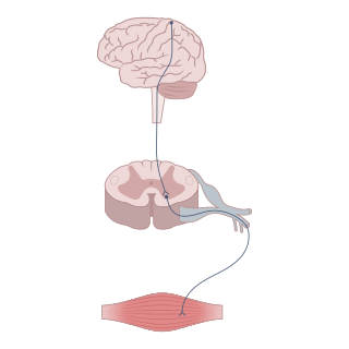
A neurotransmitter is a signaling molecule secreted by a neuron to affect another cell across a synapse. The cell receiving the signal, or target cell, may be another neuron, but could also be a gland or muscle cell.

Serotonin or 5-hydroxytryptamine (5-HT) is a monoamine neurotransmitter. Its biological function is complex, touching on diverse functions including mood, cognition, reward, learning, memory, and numerous physiological processes such as vomiting and vasoconstriction.

The raphe nuclei are a moderate-size cluster of nuclei found in the brain stem. They have 5-HT1 receptors which are coupled with Gi/Go-protein-inhibiting adenyl cyclase. They function as autoreceptors in the brain and decrease the release of serotonin. The anxiolytic drug Buspirone acts as partial agonist against these receptors. Selective serotonin reuptake inhibitor (SSRI) antidepressants are believed to act in these nuclei, as well as at their targets.

5-HT receptors, 5-hydroxytryptamine receptors, or serotonin receptors, are a group of G protein-coupled receptor and ligand-gated ion channels found in the central and peripheral nervous systems. They mediate both excitatory and inhibitory neurotransmission. The serotonin receptors are activated by the neurotransmitter serotonin, which acts as their natural ligand.

The periaqueductal gray (PAG), also known as the central gray, is a brain region that plays a critical role in autonomic function, motivated behavior and behavioural responses to threatening stimuli. PAG is also the primary control center for descending pain modulation. It has enkephalin-producing cells that suppress pain.

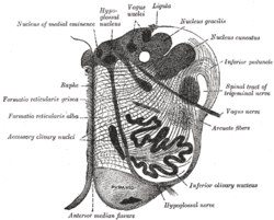
The reticular formation is a set of interconnected nuclei in the brainstem that spans from the lower end of the medulla oblongata to the upper end of the midbrain. The neurons of the reticular formation make up a complex set of neural networks in the core of the brainstem. The reticular formation is made up of a diffuse net-like formation of reticular nuclei which is not well-defined. It may be seen as being made up of all the interspersed cells in the brainstem between the more compact and named structures.
Neuromodulation is the physiological process by which a given neuron uses one or more chemicals to regulate diverse populations of neurons. Neuromodulators typically bind to metabotropic, G-protein coupled receptors (GPCRs) to initiate a second messenger signaling cascade that induces a broad, long-lasting signal. This modulation can last for hundreds of milliseconds to several minutes. Some of the effects of neuromodulators include altering intrinsic firing activity, increasing or decreasing voltage-dependent currents, altering synaptic efficacy, increasing bursting activity and reconfiguring synaptic connectivity.

The dorsal raphe nucleus is one of the raphe nuclei. It is situated in the brainstem at the midline. It has rostral and caudal subdivisions:

The median raphe nucleus(MRN), also known as the superior central nucleus, is a nucleus in the brainstem composed of polygonal, fusiform, and piriform neurons, which exists rostral to the pontine raphe nucleus. The median raphe nucleus is one of several raphe nuclei that lies on the brainstem midline. It is one of two nuclei that are situated more superior to the others. The second of these nuclei is the dorsal raphe nucleus (DRN). The MRN extends from the lower part of the dorsal raphe nucleus to an approximate position at the decussation of the superior cerebellar peduncle.
The nucleus raphe magnus (NRM) is one of the seven raphe nuclei. It is situated in the pons in the brainstem, just rostral to the nucleus raphe obscurus.

The lateral hypothalamus (LH), also called the lateral hypothalamic area (LHA), contains the primary orexinergic nucleus within the hypothalamus that widely projects throughout the nervous system; this system of neurons mediates an array of cognitive and physical processes, such as promoting feeding behavior and arousal, reducing pain perception, and regulating body temperature, digestive functions, and blood pressure, among many others. Clinically significant disorders that involve dysfunctions of the orexinergic projection system include narcolepsy, motility disorders or functional gastrointestinal disorders involving visceral hypersensitivity, and eating disorders.
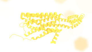
The 5-HT2C receptor is a subtype of the 5-HT2 receptor that binds the endogenous neurotransmitter serotonin (5-hydroxytryptamine, 5-HT). Like all 5-HT2 receptors, it is a G protein-coupled receptor (GPCR) that is coupled to Gq/G11 and mediates excitatory neurotransmission. HTR2C denotes the human gene encoding for the receptor, that in humans is located on the X chromosome. As males have one copy of the gene and females have one of the two copies of the gene repressed, polymorphisms at this receptor can affect the two sexes to differing extent.

The serotonin 1A receptor is a subtype of serotonin receptors, or 5-HT receptors, that binds serotonin, also known as 5-HT, a neurotransmitter. 5-HT1A is expressed in the brain, spleen, and neonatal kidney. It is a G protein-coupled receptor (GPCR), coupled to the Gi protein, and its activation in the brain mediates hyperpolarization and reduction of firing rate of the postsynaptic neuron. In humans, the serotonin 1A receptor is encoded by the HTR1A gene.

WAY-100635 is a piperazine drug and research chemical widely used in scientific studies. It was originally believed to act as a selective 5-HT1A receptor antagonist, but subsequent research showed that it also acts as potent full agonist at the D4 receptor. It is sometimes referred to as a silent antagonist at the former receptor. It is closely related to WAY-100135.

para-Chloroamphetamine (PCA), also known as 4-chloroamphetamine (4-CA), is a serotonin–norepinephrine–dopamine releasing agent (SNDRA) and serotonergic neurotoxin of the amphetamine family. It is used in scientific research in the study of the serotonin system, as a serotonin releasing agent (SRA) at lower doses to produce serotonergic effects, and as a serotonergic neurotoxin at higher doses to produce long-lasting depletions of serotonin.
Sleep onset is the transition from wakefulness into sleep. Sleep onset usually transits into non-rapid eye movement sleep but under certain circumstances it is possible to transit from wakefulness directly into rapid eye movement sleep.
Serotonergic cell groups refer to collections of neurons in the central nervous system that have been demonstrated by histochemical fluorescence to contain the neurotransmitter serotonin (5-hydroxytryptamine). Since they are for the most part localized to classical brainstem nuclei, particularly the raphe nuclei, they are more often referred to by the names of those nuclei than by the B1-9 nomenclature. These cells appear to be common across most mammals and have two main regions in which they develop; one forms in the mesencephlon and the rostral pons and the other in the medulla oblongata and the caudal pons.

LP-44 is a drug which acts as a potent and selective agonist at the 5HT7 serotonin receptor. While LP-44 is less selective than the related compound LP-12, it has been more widely used in research and has been used to show the complex role of 5-HT7 receptors in several aspects of brain function, including regulation of the sleep-wake cycle and roles in stress, learning and memory.
The raphespinal tract is a descending spinal cord tract located in the medulla oblongata. It consists of two tracts an anterior raphespinal tract, and a lateral raphespinal tract that mainly descend in the lateral funiculus. Fibers descend in the ventral portion of the lateral funiculus, mainly bilaterally to terminate in laminae I, II, and IV.
Monoamine nuclei are clusters of cells that primarily use monoamine neurotransmitters to communicate. The raphe nuclei, ventral tegmental area, and locus coeruleus have been included in texts about monoamine nuclei. These nuclei receive a variety of inputs including from other monoamines, as well as from glutaminergic, GABAergic, and substance p related pathways. The catacholaminergic pathways mainly project upwards into the cortical and limbic regions, power sparse descending axons have been observed in animals models. Both ascending and descending serotonergic pathways project from the raphe nuclei. Raphe nuclei in the obscurus, pallid us, and magnus descend into the brainstem and spinal cord, while the raphe ponds, raphe dorsals, and nucleus centralism superior projected up into the medial forebrain bundle before branching off. Monoamine nuclei have been studied in relation to major depressive disorder, with some abnormalities observed, however MAO-B levels appear to be normal during depression in these regions.