Anterior median fissure of the medulla oblongata

Last updated
Anterior median fissure of the medulla oblongata
Gray694.png
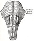
Section of the medulla oblongata at about the middle of the olive. (Anterior median fissure labeled at bottom center.)
Details
Identifiers
Latin fissura mediana anterior medullae oblongatae
NeuroNames 700
TA A14.1.04.001
FMA 83734
Anatomical terms of neuroanatomy

The anterior median fissure (ventral or ventromedian fissure) contains a fold of pia mater, and extends along the entire length of the medulla oblongata: It ends at the lower border of the pons in a small triangular expansion, termed the foramen cecum.

Pia mater delicate innermost layer of the meninges, the membranes surrounding the brain and spinal cord

Pia mater, often referred to as simply the pia, is the delicate innermost layer of the meninges, the membranes surrounding the brain and spinal cord. Pia mater is medieval Latin meaning "tender mother". The other two meningeal membranes are the dura mater and the arachnoid mater. Both the pia and arachnoid mater are derivatives of the neural crest while the dura is derived from embryonic mesoderm. The pia mater is a thin fibrous tissue that is permeable to water and small solutes. The pia mater allows blood vessels to pass through and nourish the brain. The perivascular space between blood vessels and pia mater is proposed to be part of a psuedolymphatic system for the brain. When the pia mater becomes irritated and inflamed the result is meningitis.

Medulla oblongata

The medulla oblongata is a long stem-like structure located in the brainstem. It is anterior and partially inferior to the cerebellum. It is a cone-shaped neuronal mass responsible for autonomic (involuntary) functions ranging from vomiting to sneezing. The medulla contains the cardiac, respiratory, vomiting and vasomotor centers and therefore deals with the autonomic functions of breathing, heart rate and blood pressure.

Its lower part is interrupted by bundles of fibers that cross obliquely from one side to the other, and constitute the pyramidal decussation.

Some fibers, termed the anterior external arcuate fibers, emerge from the fissure above this decussation and curve lateralward and upward over the surface of the medulla oblongata to join the inferior peduncle.

Anterior external arcuate fibers

The anterior external arcuate fibers vary as to their prominence: in some cases they form an almost continuous layer covering the medullary pyramids and olivary body, while in other cases they are barely visible on the surface.

Additional images

Reticular formation spinal trigeminal nucleus

The reticular formation is a set of interconnected nuclei that are located throughout the brainstem. The reticular formation is not anatomically well defined because it includes neurons located in different parts of the brain. The neurons of the reticular formation make up a complex set of networks in the core of the brainstem that stretch from the upper part of the midbrain to the lower part of the medulla oblongata. The reticular formation includes ascending pathways to the cortex in the ascending reticular activating system (ARAS) and descending pathways to the spinal cord via the reticulospinal tracts of the descending reticular formation.

Related Research Articles

Pons part of the brainstem

The pons is part of the brainstem, and in humans and other bipeds lies inferior to the midbrain, superior to the medulla oblongata and anterior to the cerebellum.

Brainstem posterior part of the brain, adjoining and structurally continuous with the spinal cord

The brainstem is the posterior part of the brain, continuous with the spinal cord. In the human brain the brainstem includes the midbrain, and the pons and medulla oblongata of the hindbrain. Sometimes the diencephalon, the caudal part of the forebrain, is included.

Pyramidal tracts

The pyramidal tracts include both the corticobulbar tract and the corticospinal tract. These are aggregations of efferent nerve fibers from the upper motor neurons that travel from the cerebral cortex and terminate either in the brainstem (corticobulbar) or spinal cord (corticospinal) and are involved in the control of motor functions of the body.

Medial lemniscus

The medial lemniscus, also known as Reil's band or Reil's ribbon, is a large ascending bundle of heavily myelinated axons that decussate in the brainstem, specifically in the medulla oblongata. The medial lemniscus is formed by the crossings of the internal arcuate fibers. The internal arcuate fibers are composed of axons of nucleus gracilis and nucleus cuneatus. The axons of the nucleus gracilis and nucleus cuneatus in the medial lemniscus have cell bodies that lie contralaterally.

Medial medullary syndrome

Medial medullary syndrome, also known as inferior alternating syndrome, hypoglossal alternating hemiplegia, lower alternating hemiplegia, or Dejerine syndrome, is a type of alternating hemiplegia characterized by a set of clinical features resulting from occlusion of the anterior spinal artery. This results in the infarction of medial part of the medulla oblongata.

Cuneate nucleus

One of the dorsal column nuclei, the cuneate nucleus is a wedge-shaped nucleus in the closed part of the medulla oblongata, in the brainstem. It contains cells that give rise to the cuneate tubercle, visible on the posterior aspect of the medulla. It lies laterally to the gracile nucleus and medial to the spinal trigeminal nucleus in the medulla.

Olivary body

In anatomy, the olivary bodies or simply olives are a pair of prominent oval structures in the medulla oblongata, the lower portion of the brainstem. They contain the olivary nuclei.

Precentral gyrus brain structure

The precentral gyrus is a prominent gyrus on the surface of the posterior frontal lobe of the brain. It is the site of the primary motor cortex that in humans is cytoarchitecturally defined as Brodmann area 4.

Inferior cerebellar peduncle

The upper part of the posterior district of the medulla oblongata is occupied by the inferior cerebellar peduncle, a thick rope-like strand situated between the lower part of the fourth ventricle and the roots of the glossopharyngeal and vagus nerves.

Arcuate nucleus (medulla)

In the medulla oblongata, the arcuate nucleus is a group of neurons located on the anterior surface of the medullary pyramids. These nuclei are the extension of the pontine nuclei. They receive fibers from the corticospinal tract and send their axons through the anterior external arcuate fibers and medullary striae to the cerebellum via the inferior cerebellar peduncle.

Anterior median fissure of spinal cord

The anterior median fissure of the spinal cord has an average depth of about 3 mm, but this is increased in the lower part of the spinal cord.

Anterior grey column

The anterior grey column is the front column of grey matter in the spinal cord. It is one of the three grey columns. The anterior grey column contains motor neurons that affect the skeletal muscles while the posterior grey column receives information regarding touch and sensation. The anterior grey column is the column where the cell bodies of alpha motor neurons are located.

Gracile fasciculus

The gracile fasciculus is a nerve tract in the dorsal column-medial lemniscus pathway of the spinal cord and carries information from the lower parts of the body. The gracile fasiculus is one of many ascending tracts which carry received sensory information to the brain via the spinal cord. It is also one of the dorsal columns, the other being the cuneate fasciculus.

Lateral corticospinal tract

The lateral corticospinal tract is the largest part of the corticospinal tract. It extends throughout the entire length of the spinal cord, and on transverse section appears as an oval area in front of the posterior column and medial to the posterior spinocerebellar tract.

Anterior corticospinal tract

The anterior corticospinal tract is a small bundle of descending fibers that connect the cerebral cortex to the spinal cord. Descending tracts are pathways by which motor signals are sent from upper motor neurons in the brain to lower motor neurons which then directly innervate muscle to produce movement. The anterior corticospinal tract is usually small, varying inversely in size with the lateral corticospinal tract, which is the main part of the corticospinal tract.

Internal arcuate fibers

The internal arcuate fibers are the axons of second-order sensory neurons that compose the gracile and cuneate nuclei of the medulla oblongata. These second-order neurons begin in the posterior grey column in the spinal cord. They receive input from first-order sensory neurons, which provide sensation to many areas of the body and have cell bodies in the dorsal root ganglia of the dorsal root of the spinal nerves. Upon decussation from one side of the medulla to the other, also known as the sensory decussation, they are then called the medial lemniscus.

Dorsal column nuclei

In neuroanatomy, the dorsal column nuclei are a pair of nuclei in the dorsal columns in the brainstem. The name refers collectively to the cuneate nucleus and gracile nucleus, which are present at the junction between the spinal cord and the medulla oblongata. Both nuclei contain second-order neurons of the dorsal column-medial lemniscus pathway, which carries fine touch and proprioceptive information from the body to the brain. Each nucleus has an associated nerve tract, the gracile fasciculus and the cuneate fasciculus.

Medullary pyramids (brainstem)

The medullary pyramids are paired white matter structures of the brainstem's medulla oblongata that contain motor fibers of the corticospinal and corticobulbar tracts – known together as the pyramidal tracts. The lower limit of the pyramids is marked when the fibers cross (decussate).

References

This article incorporates text in the public domain from page 767 of the 20th edition of Gray's Anatomy (1918)

The public domain consists of all the creative works to which no exclusive intellectual property rights apply. Those rights may have expired, been forfeited, expressly waived, or may be inapplicable.

<i>Grays Anatomy</i> English-language textbook of human anatomy

Gray's Anatomy is an English language textbook of human anatomy originally written by Henry Gray and illustrated by Henry Vandyke Carter. Earlier editions were called Anatomy: Descriptive and Surgical and Gray's Anatomy: Descriptive and Applied, but the book's name is commonly shortened to, and later editions are titled, Gray's Anatomy. The book is widely regarded as an extremely influential work on the subject, and has continued to be revised and republished from its initial publication in 1858 to the present day. The latest edition of the book, the 41st, was published in September 2015.