
The pons is part of the brainstem, and in humans and other bipeds lies inferior to the midbrain, superior to the medulla oblongata and anterior to the cerebellum.

The brainstem is the posterior part of the brain, continuous with the spinal cord. In the human brain the brainstem includes the midbrain, and the pons and medulla oblongata of the hindbrain. Sometimes the diencephalon, the caudal part of the forebrain, is included.

The diencephalon is a division of the forebrain, and is situated between the telencephalon and the midbrain. It consists of structures that are on either side of the third ventricle, including the thalamus, the hypothalamus, the epithalamus and the subthalamus.

The upper part of the posterior district of the medulla oblongata is occupied by the inferior cerebellar peduncle, a thick rope-like strand situated between the lower part of the fourth ventricle and the roots of the glossopharyngeal and vagus nerves.

The posterior inferior cerebellar artery (PICA), the largest branch of the vertebral artery, is one of the three main arterial blood supplies for the cerebellum, part of the brain. Occlusion of the posterior inferior cerebellar artery or one of its branches, or of the vertebral artery leads to lateral medullary syndrome also called Wallenberg syndrome.

The gracile fasciculus is a nerve tract in the dorsal column-medial lemniscus pathway of the spinal cord and carries information from the lower parts of the body. The gracile fasiculus is one of many ascending tracts which carry received sensory information to the brain via the spinal cord. It is also one of the dorsal columns, the other being the cuneate fasciculus.

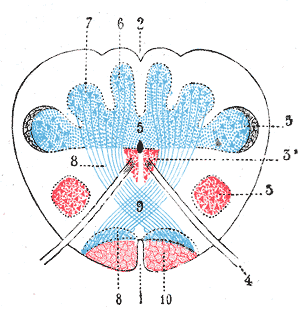
The rhomboid fossa is a rhombus-shaped depression that is the anterior part of the fourth ventricle. Its anterior wall, formed by the back of the pons and the medulla oblongata, constitutes the floor of the fourth ventricle.
In neuroanatomy, the dorsal column nuclei are a pair of nuclei in the dorsal columns in the brainstem. The name refers collectively to the cuneate nucleus and gracile nucleus, which are present at the junction between the spinal cord and the medulla oblongata. Both nuclei contain second-order neurons of the dorsal column-medial lemniscus pathway, which carries fine touch and proprioceptive information from the body to the brain. Each nucleus has an associated nerve tract, the gracile fasciculus and the cuneate fasciculus.

Winding around the inferior cerebellar peduncle in the lower part of the fourth ventricle, and crossing the area acustica and the medial eminence are a number of white strands, the medullary striae, which form a portion of the cochlear division of the vestibulocochlear nerve and disappear into the median sulcus.
In the brain, the taenia of the fourth ventricle are two narrow bands of white matter, one on either side, which complete the lower part of the roof of the fourth ventricle.
The posterior spinal artery arises from the vertebral artery in 25% of humans or the posterior inferior cerebellar artery in 75% of humans, adjacent to the medulla oblongata. It supplies the grey and white posterior columns of the spinal cord.

The dorsal nucleus of the vagus nerve is a cranial nerve nucleus for the vagus nerve in the medulla that lies ventral to the floor of the fourth ventricle. It mostly serves parasympathetic vagal functions in the gastrointestinal tract, lungs, and other thoracic and abdominal vagal innervations. The cell bodies for the preganglionic parasympathetic vagal neurons that innervate the heart reside in the nucleus ambiguus.
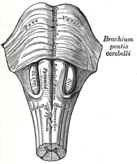
The medullary pyramids are paired white matter structures of the brainstem's medulla oblongata that contain motor fibers of the corticospinal and corticobulbar tracts – known together as the pyramidal tracts. The lower limit of the pyramids is marked when the fibers cross (decussate).
Median sulcus can refer to:
Posterior median sulcus can refer to:

The nodule, or anterior end of the inferior vermis, abuts against the roof of the fourth ventricle, and can only be distinctly seen after the cerebellum has been separated from the medulla oblongata and pons.
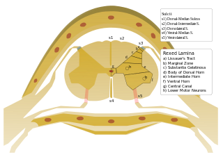
On either side of the posterior median sulcus, and at a short distance from it, the posterior nerve roots are attached along a vertical furrow named the posterolateral sulcus. The portion of the medulla spinalis which lies between this and the posterior median sulcus is named the posterior funiculus.
Median fissure may refer to:
The public domain consists of all the creative works to which no exclusive intellectual property rights apply. Those rights may have expired, been forfeited, expressly waived, or may be inapplicable.

Gray's Anatomy is an English language textbook of human anatomy originally written by Henry Gray and illustrated by Henry Vandyke Carter. Earlier editions were called Anatomy: Descriptive and Surgical, Anatomy of the Human Body and Gray's Anatomy: Descriptive and Applied, but the book's name is commonly shortened to, and later editions are titled, Gray's Anatomy. The book is widely regarded as an extremely influential work on the subject, and has continued to be revised and republished from its initial publication in 1858 to the present day. The latest edition of the book, the 41st, was published in September 2015.


